تخطيط كهربية الدماغ
EEG
مراجعة من قبل Dr John Cox, MRCGPآخر تحديث بواسطة الدكتور كولين تايدي، MRCGPLast updated 24 May 2018
يتوافق مع الإرشادات التحريرية
- تنزيلتنزيل
- مشاركة
- Language
- نقاش
- نسخة صوتية
- Add to preferred sources on Google
في هذه السلسلة:الصرعأنواع النوباتالنوبات التوترية الرمعيةالنوبات البؤريةنوبات الغيابأدوية الصرع والآثار الجانبية
تم أرشفة هذه الصفحة.
لم يتم مراجعته مؤخرًا وليس محدثًا. قد لا تعمل الروابط والمراجع الخارجية بعد الآن.
An electroencephalograph is a useful test to help diagnose epilepsy. It records the electrical activity of the brain. However, a normal result does not rule out epilepsy.
ملاحظة: المعلومات أدناه هي دليل عام فقط. قد تختلف الترتيبات وطريقة إجراء الاختبارات بين المستشفيات المختلفة. دائماً اتبع التعليمات المقدمة من طبيبك أو المستشفى المحلي.
At a glance
An electroencephalograph (EEG) records electrical signals from your brain.
It is a painless and harmless test that usually takes 20-30 minutes.
Small patches (electrodes) are placed on your scalp and connected to the EEG machine.
An EEG can help diagnose conditions like epilepsy, especially if you have had seizure-like symptoms.
You may be asked to blink or breathe deeply during the test.
A normal EEG does not rule out epilepsy, and an abnormal result does not always mean you have epilepsy.
في هذه المقالة:
Video picks for اختبارات وتحقيقات أخرى
تابع القراءة أدناه
What is an electroencephalograph?
The brain normally produces tiny electrical signals that come from the brain cells and nerves which send messages to each other. These electrical signals can be detected and recorded by the electroencephalograph (EEG) machine. The EEG test is painless and harmless. (The EEG machine records electrical signals coming from your brain - it does not put any electricity into your brain or body.)
You may be advised to have an EEG if you have had symptoms which may be due to a seizure. (Old words for seizure are convulsion or 'fit'.) An EEG test is useful in diagnosing conditions such as epilepsy. See the separate leaflet called Types of Epilepsy and Seizures.
How is the test done?
العودة إلى المحتوياتWhen you have the test, the operator will attach several small patches (electrodes) to your scalp. Wires from the electrodes are connected to the EEG machine. The machine detects and amplifies the electrical signals and records them on to a paper or computer. The test takes about 20-30 minutes. The electrodes are removed at the end of the test.
EEG machine
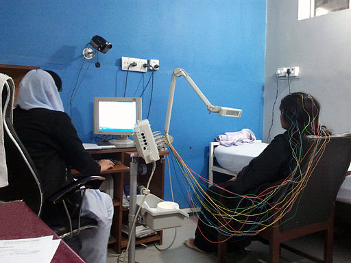
© By Baitaal (Own work), via Wikimedia Commons
For the duration of the test you may be asked to sit in a chair or lie on a couch. At some point you may be asked to blink lots of times, or to breathe deeply. These can sometimes trigger patterns of electrical activity in the brain which are associated with certain types of epilepsy.
تابع القراءة أدناه
What can the test show?
العودة إلى المحتوياتA normal ('negative') result
This shows a typical pattern of electrical activity from the brain. Most people without epilepsy, and many people with epilepsy, have a normal result. This is because an EEG only shows the electrical activity of the brain when the test is done. With many types of epilepsy, you only have abnormal electrical activity during a seizure. For the rest of the time, the pattern is normal.
An abnormal ('positive') result
This shows abnormal patterns of electrical activity. Some people with certain types of epilepsy have abnormal patterns all the time, not just when they have seizures. (Although, during a seizure the activity is even more abnormal.) For example, children with typical 'absence seizures' often have a characteristic EEG pattern which helps to confirm this type of epilepsy.
However, a small number of people who never have seizures and who don't have epilepsy, have some abnormal patterns of electrical activity in the brain.
Therefore, if you have symptoms which are thought to be seizures, an abnormal EEG means that the diagnosis is likely to be epilepsy. However, a normal result does not rule out epilepsy, and an abnormal result does not necessarily mean that you have epilepsy.
Children and electroencephalography
العودة إلى المحتوياتThe interpretation of a child's EEG recording is more difficult. This is because the EEG changes during childhood. An adult pattern is usually developed by the age of 15 years. As the EEG pattern in infants and children can vary considerably, careful interpretation of the test is necessary.
تابع القراءة أدناه
Some specialised types of electroencephalograph test
العودة إلى المحتوياتStrobe lighting
In some cases, a strobe light may be used during an EEG test. This aims to detect if this alters the electrical pattern in the brain. (Usually it does not. However, a small number of people have seizures triggered by flickering or strobe lights and so this may help to identify these people.)
Sleep EEG
In this test, an EEG is performed while you are sleeping. This is usually carried out when you are in hospital. You may need to have one of these if your seizures happen when you are asleep or when you are tired.
Sleep-deprived EEG
There may be a better chance of detecting abnormal brain activity after a period of time when you are deprived of sleep. Therefore, sometimes the EEG test is done after you have stayed awake for all or most of the night. It is done in the same way as the normal test but with you asleep - after the period of 'sleep deprivation'.
Ambulatory EEG
This may be advised in cases where the diagnosis is not clear. It uses a portable EEG machine which records the brain's electrical activity when you are going about your normal activities. The electrodes can usually be hidden under your hair. The wires are connected to a small machine which you wear on a belt (a bit like wearing an mp3 player).
You may be asked to keep a diary when you have meals, go to sleep, and have any symptoms which may be a seizure. The patterns on the EEG can be analysed to see if they change when symptoms occur. It may help to confirm whether certain symptoms are due to seizures.
Video-telemetry
Where there is doubt about a diagnosis of epilepsy, or where the type of seizures someone experiences is unclear, video-telemetry can be helpful. This is a test that uses a video camera linked to an EEG machine.
The camera will visually record your movements and, at the same time, the EEG machine will record your brainwave pattern. Both the video and EEG are stored on to a computer so that they can be reviewed once the test is finished.
The doctor will be able to see any seizures that you may have had, as well as any changes in your EEG at that time. The test is often carried out over a number of days in order to increase the chances of recording one of your seizures.
What should I do to prepare for an electroencephalograph?
العودة إلى المحتوياتYour doctor should give you instructions about what you need to do to prepare for an EEG. Commonly there is no preparation necessary. You should not stop any medication you take for seizures unless advised to by your doctor.
Patient picks for اختبارات وتحقيقات أخرى

الفحوصات والتحقيقات
Audiology
If you or a family member suspect that you have a hearing problem, then an audiologist will usually provide an assessment and also may be involved in providing any treatment needed. Certain conditions causing balance difficulties or dizziness can arise due to problems in the ear and audiologists can undertake specialist tests to investigate these problems as well.
بواسطة الدكتورة سورانجي مينديس، MRCGP

الفحوصات والتحقيقات
Fine-needle aspiration
In fine-needle aspiration a thin, hollow needle is used to take a sample of cells from an organ or lump under the skin. The cells are then analysed under a microscope. Note: the information below is a general guide only. The arrangements and the way tests are performed, may vary between different hospitals. Always follow the instructions given by your doctor or local hospital.
by Dr Roger Henderson, MBBS
الأسئلة الشائعة
Will I feel anything when the electrical signals are detected by the EEG machine?
No, the EEG test is painless and harmless. The machine records the electrical signals naturally produced by your brain; it does not send any electricity into your brain or body.
What specifically happens during the test to try and trigger brain activity?
During the test, you might be asked to blink many times or to breathe deeply. These actions can sometimes help to trigger specific patterns of electrical activity in your brain that are linked to certain types of epilepsy.
If my initial EEG is normal, does that mean I definitely don't have epilepsy?
Not necessarily. A normal (negative) result indicates a typical pattern of electrical activity at the time of the test. However, with many types of epilepsy, abnormal electrical activity only occurs during a seizure. For the rest of the time, the brain's electrical pattern can appear normal. Therefore, a normal result doesn't rule out epilepsy.
What if my EEG shows abnormal patterns, but I don't have seizures?
It is possible for a small number of people who never have seizures and do not have epilepsy to show some abnormal electrical patterns in their brain during an EEG. Therefore, an abnormal result doesn't automatically mean you have epilepsy.
How does an ambulatory EEG work if I'm moving around?
An ambulatory EEG involves a portable machine that records your brain's electrical activity as you go about your daily activities. The electrodes are usually hidden under your hair, and they are connected to a small machine worn on a belt. You might be asked to keep a diary of your meals, sleep, and any potential seizure symptoms to help analyse the patterns recorded.
Why would a video-telemetry test be recommended?
Video-telemetry is useful when there is uncertainty about an epilepsy diagnosis or if the specific type of seizures someone experiences is unclear. This test combines a video camera, which records your movements, with an EEG machine, which records your brainwave pattern simultaneously. This allows doctors to match visual seizure events with changes in your brain's electrical activity. It's often conducted over several days to increase the chance of recording a seizure.
قراءة إضافية ومراجع
- Epilepsies: diagnosis and management; NICE Clinical Guideline (January 2012)
- تشخيص وإدارة الصرع لدى البالغين; الشبكة الاسكتلندية للإرشادات المشتركة بين الكليات - SIGN (2015 - تم التحديث 2018)
- الصرع; NICE CKS, March 2018 (UK access only)
تابع القراءة أدناه
About the authorView full bio

الدكتور كولين تايدي، MRCGP
General Practitioner, Medical Author
MBBS, MRCGP, MRCP (Paediatrics), DCH
Dr Colin Tidy is an NHS Doctor, based in Oxfordshire.
About the reviewerView full bio

Dr John Cox, MRCGP
MA, MB, B Chir, DCH, DRCOG, MRCP (UK), MRCGP
Dr John Cox worked as a Medical Registrar in the UK and New Zealand and as a locum Physician in New Zealand.
تاريخ المقال
تمت كتابة المعلومات على هذه الصفحة ومراجعتها من قبل أطباء مؤهلين.
24 May 2018 | أحدث إصدار

اسأل، شارك، تواصل.
تصفح المناقشات، اطرح الأسئلة، وشارك التجارب عبر مئات المواضيع الصحية.

هل تشعر بتوعك؟
قم بتقييم أعراضك عبر الإنترنت مجانًا
اشترك في النشرة الإخبارية للمرضى
جرعتك الأسبوعية من النصائح الصحية الواضحة والموثوقة - مكتوبة لمساعدتك على الشعور بالاطلاع والثقة والتحكم.
By subscribing you accept our سياسة الخصوصية. يمكنك إلغاء الاشتراك في أي وقت. نحن لا نبيع بياناتك أبدًا.