Club foot
Congenital talipes equinovarus
Peer reviewed by Dr Hayley Willacy, FRCGP Last updated by Dr Doug McKechnie, MRCGPLast updated 2 Jan 2024
Meets Patient’s editorial guidelines
- DownloadDownload
- Share
- Language
- Discussion
- Audio Version
- Add to preferred sources on Google
Club foot (also called talipes equinovarus is a deformity of the foot and ankle that a baby can be born with.
It is not clear exactly what causes club foot. In most cases, it is diagnosed by the typical appearance of a baby's foot after they are born.
The Ponseti method is a widely used treatment for club foot. This treatment gives good results for most children. If it doesn't work, surgery can help.
At a glance
Club foot is a foot and ankle deformity that a baby is born with.
It causes the foot to point downwards and turn inwards, making it appear short and wide.
Around 1 in 1,000 babies in the UK are born with club foot.
The Ponseti method is the main treatment, involving gentle foot manipulation and plaster casts.
A minor operation called an Achilles tenotomy may also be needed.
Babies typically wear a brace (special boots and a bar) until they are four years old.
Early treatment usually leads to good correction of the deformity.
What is club foot?
Club foot
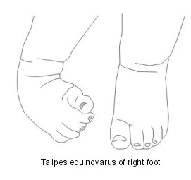
Club foot, also known as talipes. It is a deformity of the foot and ankle that a baby can be born with.
If a baby has club foot, their foot points downwards at their ankle and the heel of their foot is turned inwards.
The middle section of their foot is also twisted inwards so their foot appears quite short and wide. It cannot be gently moved into a normal foot position.
The baby's foot is kept in this position because the Achilles tendon at the back of the baby's heel is very tight and the tendons on the inside of their leg have become shortened.
If nothing is done to correct the problem, as the baby learns to stand, they will not be able to put the sole of their foot flat on the ground.
Some babies hold their foot in a position that can look as if they have club foot but, in fact, their foot can be moved easily into a normal position. These babies do not have true club foot.
In about half of babies born with club foot, both feet are affected. 'Talipes' means the ankle and foot; 'equinovarus' refers to the position that the foot is in (see below). Club foot is a congenital condition, meaning that you are born with it.
What causes club foot?
In some cases the position of the foot is due to the way the baby was lying in the womb. The deformity can be easily corrected by a series of gentle stretches as advised by a physiotherapist. This is called positional talipes.
If you have had a baby born with club foot, there is about a 3-4 in 100 chance that a brother or sister born after them will also have the condition.
Babies born to a parent who has club foot also have an increased risk of being born with the deformity themselves. If both parents have club foot, this risk is higher. Club foot may also have something to do with the position of the baby's foot when the baby is in the womb.
In most cases (around 4 out of 5), the baby has no other problems apart from the club foot. However, in around 1 in 5 babies who are born with club foot, there is also another problem. These problems may include:
Spina bifida - a condition where the bones of the spine don't form properly, which can lead to damage to the nerves of the spine.
Cerebral palsy - a general term that describes a group of conditions that cause movement problems.
Arthrogryposis - a condition where a child has curved and stiff joints and abnormal muscle development.
Structural talipes
Sometimes the club foot cannot be corrected easily. The muscles and ligaments may be very tight and in more severe cases there may be some bony abnormality. This is called structural talipes.
It is not clear exactly why structural talipes develops. It is thought that there may be genetic risk factors involved.
How common is club foot?
Club foot is a fairly common problem. It is one of the most common deformities that a baby can be born with. About 1 in 1,000 babies born in the UK have club foot.
About twice as many boys as girls are born with club foot and it can affect both feet.
How is club foot diagnosed?
Club foot was previously only diagnosed after a baby is born. However, as the technology of ultrasound scanning during pregnancy improves, increasingly, club foot is being detected during scanning before a baby is born.
All babies in the UK are routinely examined and checked over by a doctor shortly after they are born. The doctor will look for club foot, as well as other problems that the baby may be born with. If the baby has club foot it is usually noticed during this check. Investigations such as X-rays are not usually needed to confirm the diagnosis.
Some babies with club foot have milder foot deformity than others. If a baby is diagnosed with club foot, a specialist (usually an orthopaedic surgeon) will often use a grading system to grade the severity.
A common grading system that is used is called the Pirani score. With this grading system, a grade from 0 to 6 is given. The higher the grade, the greater the degree of foot deformity.
What is the treatment for club foot?
Ponseti method
The Ponseti method is now the preferred treatment by orthopaedic surgeons throughout the world. Major surgery used to be common; however, medical research has shown that the Ponseti method gives better long-term results for most children.
This method involves the specialist gently manipulating (holding, stretching and moving) the child's foot with their hands, into a position in which the foot deformity is put right (corrected) as much as possible. This is not painful or uncomfortable for the child.
Once in this position, a plaster cast is put on to hold the child's foot in position. This plaster cast usually goes all the way from the child's toes to their groin area.
After one week, the plaster cast is removed, the child's foot is manipulated again and a plaster cast is put back on with the child's foot in the new position. After another week, this procedure is repeated.
As each week goes by, usually the child's foot is able to be moved into a position that becomes closer and closer to a normal foot position. After around six weeks of repeated manipulation and plaster casting of the foot, there is usually good progress and the foot position has improved.
Achilles tenotomy
At this stage, a small operation is suggested for most children, called an Achilles tenotomy. This involves releasing the tight Achilles tendon at the back of the foot, using a small cut so that the heel can drop down. It is a minor operation and it can usually be done with just a local anaesthetic.
After this, their foot is put in a final plaster cast, usually for three weeks. The child will then need to wear a brace (some special boots that are connected together with a bar). They will need to wear these for 23 hours a day for three months. After this they generally just need to wear the brace at night or during sleep periods until they are 4 years old.
It is really important for the child to continue to wear their 'boots and bar' as the specialist advises. If the boots and bar are not worn as advised, there is a chance that club foot can come back.
It is important that a baby who has club foot be referred to see a doctor specialised in treating this problem as soon as possible after birth. The sooner Ponseti method treatment is started, in general, the easier the correction of the foot deformity should be.
Other methods
Other treatment methods are available. One example is the French functional method, which involves daily manipulation as well as immobilisation with adhesive bandages and pads.
Kite technique
The Kite technique was widely practised until the emergence of the Ponseti technique. The Kite technique involves long leg plaster casts (toe to groin) with manipulation around the calcaneo‐cuboid joint in the foot. Casting may continue for up to two years, with more than half of cases requiring major surgical intervention.
Minor surgery
The treatment of club foot does not usually need surgery, and surgical options are reserved for correction of any remaining deformity. Minor surgical interventions may occasionally include release of the Achilles tendon (Achilles tenotomy), moving a tendon in the foot (tibialis anterior tendon transfer) or lengthening of the Achilles tendon.
Other treatments include the use of an external brace (fixator device) and botulinum toxin injections.
What is the outlook for club foot?
The Ponseti method works well to correct the foot deformity for most children with club foot. For between 8 and 9 out of 10 children, the deformity will be corrected.
However, in a small number of children, it does not correct the deformity and more major surgery may be needed.
Children who have other problems as well as club foot, such as those discussed above, are more likely to need surgery.
Dr Mary Lowth is an author or the original author of this leaflet.
Patient picks for Development
Children's health
Developmental dysplasia of the hip
Developmental dysplasia of the hip is a problem with the way that the hip joint develops. It is usually present from birth although may develop later. It is more common in girls. When developmental dysplasia of the hip is diagnosed and treated early in a young baby, the outcome is usually excellent. If treatment is delayed, the treatment is more complex and has less chance of being successful.
by Dr Jacqueline Payne, FRCGP
Children's health
Duchenne muscular dystrophy
Duchenne muscular dystrophy is a condition which causes progressive muscle weakness. It starts in childhood and may be noticed when a child has difficulty standing up, climbing or running. It is a genetic condition and can be inherited. It usually affects only boys, although girls may carry the Duchenne gene. Boys with Duchenne muscular dystrophy should have regular reviews and physiotherapy from childhood. They are likely to need increasing help and treatments.
by Dr Philippa Vincent, MRCGP
Frequently asked questions
What is 'positional talipes'?
Positional talipes is a type of club foot where the foot's position is due to how the baby was lying in the womb. This type of deformity can often be corrected with a series of gentle stretches, as advised by a physiotherapist.
What is 'structural talipes'?
Structural talipes refers to cases where the club foot cannot be easily corrected. In these situations, the muscles and ligaments may be very tight, and there might be some bony abnormalities. The exact cause of structural talipes is not fully understood, but it is thought that genetic factors might play a role.
How long does the Ponseti method treatment take?
The initial phase of the Ponseti method involves weekly plaster cast changes for about six weeks. After this, a minor operation called an Achilles tenotomy is usually performed, followed by a final plaster cast for three weeks. Following cast removal, the child will need to wear a special brace (boots connected by a bar) for 23 hours a day for three months, and then only at night or during sleep until they are 4 years old.
Are there any alternative treatments if the Ponseti method isn't suitable or doesn't work?
Yes, other treatment methods are available. One example is the French functional method, which involves daily manipulation and immobilisation with adhesive bandages and pads. If the current treatments are not sufficient, minor surgical interventions might be considered to correct any remaining deformity.
Can club foot return even after successful treatment?
Yes, there is a chance that club foot can return if the 'boots and bar' brace, which is a crucial part of the treatment, is not worn exactly as advised by the specialist. It is very important to follow the wearing schedule to prevent recurrence.
Why is early diagnosis and treatment important for club foot?
Early diagnosis and treatment are important because the sooner the Ponseti method treatment is started after birth, the easier the correction of the foot deformity generally is. This typically leads to better long-term results for the child.
Further reading and references
- Club Foot and the Ponseti Method; Ponseti International
- Bina S, Pacey V, Barnes EH, et al; Interventions for congenital talipes equinovarus (clubfoot). Cochrane Database Syst Rev. 2020 May 15;5:CD008602. doi: 10.1002/14651858.CD008602.pub4.
- Pavone V, Chisari E, Vescio A, et al; The etiology of idiopathic congenital talipes equinovarus: a systematic review. J Orthop Surg Res. 2018 Aug 22;13(1):206. doi: 10.1186/s13018-018-0913-z.
- Mustari MN, Faruk M, Bausat A, et al; Congenital talipes equinovarus: A literature review. Ann Med Surg (Lond). 2022 Aug 18;81:104394. doi: 10.1016/j.amsu.2022.104394. eCollection 2022 Sep.
- Gelfer Y, Blanco J, Trees A, et al; Attaining a British consensus statement on managing idiopathic congenital talipes equinovarus (CTEV) through a Delphi process: a study protocol. BMJ Open. 2021 Sep 2;11(9):e049212. doi: 10.1136/bmjopen-2021-049212.
About the authorView full bio

Dr Michelle Wright, MRCGP
General Practitioner, Medical Author
MB, ChB, MRCGP, DCH, DRCOG
Dr Michelle Wright qualified in 1997 in the UK and worked as a GP in London before moving to Switzerland. She has been an author with EMIS since 2007.
About the reviewerView full bio

Dr Hayley Willacy, FRCGP
General Practitioner, Medical Author
MBChB (1992), DRCOG, DFFP, MRCOG (Part 1) MRCGP (2007), DFSRH (2013), MSc - medical education (2020)
Dr Hayley Willacy was an NHS GP working in northwest England, who retired from clinical practice in 2022 after 30 years.
Article history
Article also available in English, German, Spanish, French, Italian, Portuguese, Hindi, Hebrew, Arabic, and Swedish.
Next review due: 1 Jan 2027
2 Jan 2024 | Latest version
27 Jan 2011 | Originally published
Authored by:
Dr Michelle Wright, MRCGP
Join the conversation
Share your thoughts or experiences with others in our patient communities.

Ask, share, connect.
Browse discussions, ask questions, and share experiences across hundreds of health topics.

Feeling unwell?
Assess your symptoms online for free
Sign up to the Patient newsletter
Your weekly dose of clear, trustworthy health advice - written to help you feel informed, confident and in control.
By subscribing you accept our Privacy Policy. You can unsubscribe at any time. We never sell your data.
More in children's health
- Adrenarche
- Albinism
- Alpha-1 antitrypsin deficiency
- Congenital heart disease
- Croup
- Duchenne muscular dystrophy
- Feeding your toddler
- Glue ear
- Hirschsprung's disease
- HPV vaccine
- Klinefelter's syndrome
- Marfan syndrome
- Meningococcal infection
- Mesenteric adenitis
- Nausea and vomiting
- Pyloric stenosis
- Restricted growth
- Rhabdomyosarcoma
- Teething