نزيف تحت العنكبوتية
مراجعة من قبل الدكتور كريشنا فاخاريا، MRCGPآخر تحديث بواسطة الدكتور كولين تايدي، MRCGPآخر تحديث ١٣ ديسمبر ٢٠٢٢
يتوافق مع الإرشادات التحريرية
- تنزيلتنزيل
- مشاركة
- Language
- نقاش
- Ask Patient
- نسخة صوتية
- أضف إلى المصادر المفضلة على جوجل
A subarachnoid haemorrhage (SAH) is a form of stroke in which bleeding occurs into the fluid-filled space surrounding the brain (called the subarachnoid space). It is usually caused by the rupture of a small berry-like swelling (called a berry aneurysm) on one of the arteries in the brain.
SAH is a medical emergency. The risk of a further bleed is greatest during the first 24 hours from when the symptoms started.
نظرة سريعة
النزيف تحت العنكبوتية هو نزيف حول الدماغ، عادةً بسبب تمدد الأوعية الدموية المنفجر.
العارض الأكثر تميزًا هو صداع مفاجئ، انفجاري، وشديد للغاية.
يمكن أن تشمل الأعراض الأخرى القيء أو النوبات أو تغير الوعي.
إذا كنت تعاني من صداع شديد ومفاجئ بشكل غير عادي مع قيء، فاطلب المشورة الطبية.
التدخين، تناول الكحول بكميات كبيرة، وارتفاع ضغط الدم يزيد من الخطر.
تهدف المعالجة إلى وقف النزيف الإضافي ومنع إصابة الدماغ.
ما هو النزيف تحت العنكبوتية؟
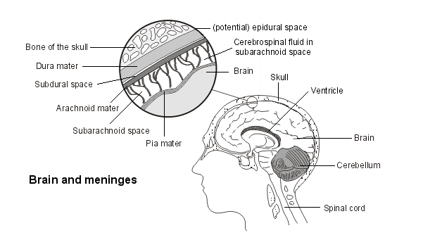
A subarachnoid haemorrhage (SAH) occurs when there is bleeding around the brain into the subarachnoid space. This is a space between two of the protective layers which surround the brain. These are the pia mater, which covers the brain directly, and the arachnoid mater, which is separated from the pia mater by a space filled with fluid. This fluid, the cerebrospinal fluid, is there to cushion and protect the brain from injury.
The bleeding usually comes from a burst (ruptured) berry aneurysm, most commonly at the back of the brain. Berry aneurysms are small berry-shaped swellings on the arteries which supply the brain, usually at a junction where an artery divides into two. These are relatively common in the general population, particularly in older patients, although most never rupture.
Brain and meninges
How serious is a subarachnoid haemorrhage?
Subarachnoid haemorrhage (SAH) is very serious. More than 1 in 4 people with a subarachnoid haemorrhage will die within the first six months after the subarachnoid haemorrhage. Of the survivors, about 1 in 3 are unable to live independently.
Who is affected by subarachnoid haemorrhage?
SAH usually affects adults under the age of 60 years, with the average age of affected patients around 50 years. Children are not usually affected.
Women are affected slightly more often than men.
Patients of African descent are slightly more commonly affected than patients of white descent.
The condition is also more common in Finland and Japan, where the rates are around three times those in Europe.
Patients who smoke, use excessive alcohol or have high blood pressure are at greater risk.
First-degree relatives of people who have had an SAH are at greater risk.
How common is subarachnoid haemorrhage?
Around 8 people out of every 100,000 per year will have an SAH.
SAH causes around 6 out of 100 of all strokes.
How does subarachnoid haemorrhage occur?
More than 8 out of 10 cases of SAH are caused by the bursting (rupture) of berry aneurysms. About 1 in every 25 adults will have one or more berry aneurysms. However, an SAH rupturing is a relatively rare event.
Larger berry aneurysms (bigger than 7 mm) are more likely to rupture (smaller ones can grow larger over time). Rupture is more likely to occur if the elastic tissue in the walls of the berry aneurysm is weakened or strained by:
ارتفاع ضغط الدم غير المسيطر عليه.
High alcohol use.
التدخين.
Cocaine use.
Certain inherited conditions which cause weakening of the blood vessels. These include autosomal dominant polycystic kidney disease (ADPKD). These conditions cause only 1-2 in every 100 cases of SAH.
Other causes of SAH, accounting for fewer than 2 out of every 10 cases, include:
Head and neck trauma, when bleeding from injury to the brain or the structures around it can track into the subarachnoid space.
Abnormal blood vessels such as those that may be present in a brain tumour.
Brain infection such as التهاب الدماغ.
Vasculitis, when blood vessels inside the brain become swollen and inflamed. This can occur either due to infection or to conditions affecting the immune system.
أعراض النزيف تحت العنكبوتية
صداع
The most characteristic feature is a sudden explosive headache.
The headache is severe, often described as the most severe headache ever experienced. It is sometimes referred to as a thunderclap headache. However, the term thunderclap headache is more commonly used for some other types of sudden headache, including صداع جنسي أولي.
Usually the headache lasts for a week or two. It is usually a dull, all-over headache. It may occasionally last only a few seconds or even less. The person may even look round and accuse someone of hitting him or her on the back of the head.
About 1 in 10 sudden explosive headaches are found to be due to SAH.
If you have an episode of sudden severe headache - coming on in minutes or less and being worse than headaches you have normally experienced - seek medical advice at once.
الشعور بالغثيان (التقيؤ)
Vomiting is very common in SAH. Vomiting also occurs with migraine and with other severe headaches. However, if you have an unusually severe sudden headache with vomiting you should seek medical advice.
Seizures and altered consciousness
A few people with SAH have a seizure due to irritation of the brain by the bleeding. Confusion or altered consciousness may also develop. If someone is unconscious they cannot tell you they have a headache. If someone complains of a severe headache then becomes unexpectedly drowsy, make sure that medical attendants are aware that the headache came first, as this suggests SAH.
Are there warning symptoms for subarachnoid haemorrhage?
Some people with SAH will have had some warning signs in the days or hours before the SAH occurs. The most common are:
Brief sudden headaches (the most common symptom).
دوار.
Eye pain.
Double vision or lost vision.
Loss of sensation or movement.
These symptoms may be caused by early small leaks of blood from the aneurysm as it starts to burst (rupture). However, they may not last long, and of course they may not be recognised as possibly serious by doctors or patients.
Subarachnoid haemorrhage CT scan
If a subarachnoid haemorrhage is suspected then a التصوير المقطعي المحوسب (CT) should be performed as soon as possible. This will nearly always confirm the diagnosis.
Other investigations may include cerebral angiography. A lumbar puncture (spinal tap) may be needed if the CT scan is normal but a subarachnoid haemorrhage is still suspected.
Subarachnoid haemorrhage treatment
Treatment of SAH
Initial treatment is aimed at:
Stabilising the person.
Preventing further bleeding
Preventing further brain injury.
Medicines are used - for example, to:
Improve blood supply to damaged parts of the brain.
Prevent seizures which can worsen brain damage.
Surgery is used in some patients:
People with SAH are usually referred to a specialist neurosurgical unit.
Treatment usually consists of blocking off the aneurysm, either:
Through blocking it from the inside by using a special platinum coil (endovascular coiling); أو
Through clipping it closed (neurosurgical clipping).
Patients need to be monitored for some time to avoid complications. These are fluid on the brain (hydrocephalus), further shortage of oxygen to the brain and low blood pressure.
Long-term management of SAH
This is aimed at:
Reducing risk of recurrence - treating high blood pressure, reducing alcohol intake and, if the person smokes, stopping smoking.
Specialist rehabilitation service for disabilities.
What is the outlook for patients with subarachnoid haemorrhage?
About 1 in 4 people die within the first six months after a subarachnoid haemorrhage. About 1 in 8 people die before reaching hospital. However more than 8 out of every 10 people admitted to a neurosurgical unit with a confirmed subarachnoid haemorrhage will survive.
Improvement will continue for about 18 months after the SAH.
Most affected people will have some impairment but some do well.
The things which increase the chance of doing better are:
A smaller bleed.
A younger age.
Remaining conscious at the time of the SAH.
About 1 in 2 people report changes with memory, mood or speech one year after a subarachnoid haemorrhage.
Can subarachnoid haemorrhage be prevented?
It is not known why some people with berry aneurysms in the brain go on to develop SAH. However, it is known that certain avoidable risk factors make it more likely:
ارتفاع ضغط الدم
التدخين
تناول الكحول بكميات مفرطة
Cocaine use
Avoiding these risk factors is the first way to reduce the risk of SAH.
The factors that affect the health of the walls of your brain's (cerebral) blood vessels are the same factors that affect your risk of heart disease (cardiovascular risk). Therefore, lifestyle measures which generally reduce cardiovascular risk will help prevent SAH. These include:
نظام غذائي صحي, following a five-a-day healthy eating plan.
Keeping a healthy مؤشر كتلة الجسم (BMI), which will help prevent SAH.
What should I do if I know I have an aneurysm?
Berry aneurysms are common. They are present in around 4 in every 100 adults, being more common with greater age. They are probably the result of wear and tear on the cerebral arteries at their point of greatest weakness - the point where they divide into two.
Aneurysms less than 7 mm in diameter tend not to burst (rupture). If you have an aneurysm which is less than 7 mm in size, you may be advised that, as long as you keep your blood pressure well controlled and maintain a healthy lifestyle, your risk of SAH is no greater than that of the general population. The decision is more difficult if your aneurysm is larger than 7 mm, or you have several aneurysms. You might be offered repeat scanning, at intervals, to see if your aneurysm is growing, and to review the decision.
The alternative is to clip or block the aneurysm, even whilst it is small. The risks of surgery are significant, particularly if you are older. The risk of rupture also increases with age. These are not easy decisions.
Incidentally found aneurysms
Because they are common, berry aneurysms are often found incidentally. That is, you are found to have a berry aneurysm when having a CT scan for another reason. If this happens you will need specialist advice as to whether you need to consider operative treatment for your aneurysm. Many factors will be taken into account when advising you, including:
عمرك.
The number of aneurysms you have.
Where the aneurysms are.
How large they are.
Any underlying conditions that might put you at greater risk.
People who have had an SAH and who have unruptured aneurysms
Those who have already had an SAH are usually offered treatment for other aneurysms, which is again an option between blocking the aneurysm from the inside using a platinum coil (endovascular coiling) or clipping it closed (neurosurgical clipping).
Screening for aneurysms
Routine testing to check for aneurysms in relatives has not been shown to be of any benefit. Any testing is based on an assessment of each relative's own risk and is usually limited to people with at least two first-degree relatives (father, mother, sister or brother) who have had a subarachnoid haemorrhage.
الدكتورة ماري لوث هي مؤلفة أو المؤلفة الأصلية لهذه النشرة.
اختيارات المرضى لـ إصابة في الرأس
الدماغ والأعصاب
ورم دموي فوق الجافية
الورم الدموي خارج الجافية هو تجمع للدم في الفراغ 'المحتمل' بين الجمجمة والبطانة الواقية الخارجية التي تغطي الدماغ (الأم الجافية). يحدث عادة بسبب إصابة في الرأس. إنه حالة خطيرة ويتطلب علاجًا طارئًا، غالبًا في شكل عملية جراحية لإزالة الورم الدموي.
بقلم الدكتورة توني هازيل، FRCGP
الدماغ والأعصاب
ارتجاج في المخ
الارتجاج هو إصابة دماغية رضية خفيفة يمكن أن تحدث بعد ضربة على الرأس أو هزة مفاجئة وشديدة.
بقلم الدكتورة فيليبا فينسنت، MRCGP
الأسئلة الشائعة
ما هو الفرق بين النزيف تحت العنكبوتية وأنواع السكتات الدماغية الأخرى؟
النزيف تحت العنكبوتية (SAH) هو نوع محدد من النزيف حول الدماغ. على الرغم من أنه يُصنف كجلطة دماغية، إلا أنه يختلف عن الجلطات الدماغية الإقفارية الأكثر شيوعًا (التي تحدث بسبب انسداد في إمداد الدم إلى الدماغ). يشكل النزيف تحت العنكبوتية حوالي 6 من كل 100 جلطة دماغية.
إذا شعرت بصداع مفاجئ وشديد، هل يعني ذلك دائمًا أنني أعاني من نزيف تحت العنكبوتية؟
لا، ليس دائمًا. في حين أن النزيف تحت العنكبوتية هو سبب شائع للصداع المفاجئ – وهو صداع مفاجئ، انفجاري، وشديد – فإن هذا المصطلح يُستخدم أيضًا لأنواع أخرى من الصداع المفاجئ. ومع ذلك، إذا شعرت بصداع مفاجئ، يجب عليك استشارة الطبيب فورًا.
لماذا تميل تمددات الأوعية الدموية التوتية إلى التمزق في الجزء الخلفي من الدماغ؟
تذكر المقالة أن تمدد الأوعية الدموية التوتي غالبًا ما يتمزق في الجزء الخلفي من الدماغ. يحدث هذا عادةً عند نقطة تقاطع حيث ينقسم الشريان إلى اثنين، وهي نقاط ضعف في الأوعية الدموية.
ما هي فرص علاج نزيف تحت العنكبوتية بنجاح إذا وصلت إلى المستشفى؟
أكثر من 8 من كل 10 أشخاص يتم إدخالهم إلى وحدة جراحة الأعصاب مع نزيف تحت العنكبوتية مؤكد سوف ينجون. يركز العلاج في البداية على استقرار حالة الشخص، ومنع المزيد من النزيف، ومنع إصابة الدماغ بمزيد من الضرر.
هل يمكنني منع تكون تمدد الأوعية الدموية التوتي؟
لا يُعرف سبب تطور بعض الأشخاص لتمدد الأوعية الدموية التوتية. يُعتقد أنها نتيجة 'التآكل والتمزق' في الشرايين الدماغية، خاصة في الأماكن التي تنقسم فيها. ومع ذلك، يمكنك اتخاذ خطوات لتقليل خطر تمزق تمدد الأوعية الدموية التوتية (مما يسبب نزيف تحت العنكبوتية) عن طريق تجنب عوامل الخطر مثل ارتفاع ضغط الدم، التدخين، تناول الكحول المفرط، واستخدام الكوكايين. كما أن نمط الحياة الصحي، بما في ذلك ممارسة الرياضة بانتظام واتباع نظام غذائي جيد، يساعد أيضًا.
بعد حدوث نزيف تحت العنكبوتية، كم من الوقت يستغرق عملية التعافي وما هي الآثار طويلة الأمد التي قد أتوقعها؟
يمكن أن يستمر التحسن بعد حدوث نزيف تحت العنكبوتية لمدة تصل إلى حوالي 18 شهرًا. بينما يحقق بعض الأشخاص نتائج جيدة جدًا، فإن معظم الأفراد المتأثرين سيعانون من بعض الإعاقات. حوالي نصف الأشخاص يبلغون عن تغييرات في الذاكرة أو المزاج أو الكلام بعد مرور عام على حدوث النزيف تحت العنكبوتية.
قراءة إضافية ومراجع
- Ziu E, Mesfin FB; Subarachnoid Hemorrhage. StatPearls, 2022.
- Etminan N, Chang HS, Hackenberg K, et al; معدل حدوث النزيف تحت العنكبوتية الناتج عن تمدد الأوعية الدموية في جميع أنحاء العالم وفقًا للمنطقة والفترة الزمنية وضغط الدم وانتشار التدخين في السكان: مراجعة منهجية وتحليل تلوي. JAMA Neurol. 1 مايو 2019;76(5):588-597. doi: 10.1001/jamaneurol.2019.0006.
- السكتة الدماغية والنوبة الإقفارية العابرة; NICE CKS، أغسطس 2020 (الوصول متاح فقط في المملكة المتحدة)
- تقييم - صداع; NICE CKS، مارس 2022 (الوصول متاح فقط في المملكة المتحدة)
- النزيف تحت العنكبوتية الناجم عن تمزق تمدد الأوعية الدموية: التشخيص والعلاج; إرشادات NICE (نوفمبر 2022)
عن المؤلفعرض السيرة الذاتية الكاملة
تم التحديث بواسطة

الدكتور كولين تايدي، MRCGP
طبيب عام، مؤلف طبي
MBBS, MRCGP, MRCP (Paediatrics), DCH
الدكتور كولين تايدي هو طبيب في هيئة الخدمات الصحية الوطنية، ويعمل في أوكسفوردشاير.
كتبه

الدكتورة ماري إليزابيث لوث، زميلة الكلية الملكية للأطباء العامين
MA (Cantab), MB, BChir, DFFP, DRCOG, PG Cert, Med Ed, FRCGP, MA (London)
Dr Mary Lowth qualified in Cambridge in 1988 and has been a GP and expedition doctor for 30 years, a medical educator and MRCGP examiner for 20 years and a law and ethics academic for 10 years. As a GP her special interests are paediatrics, ethics and womens health, and sexual health medicine.
حول المراجععرض السيرة الذاتية الكاملة

الدكتور كريشنا فاخاريا، MRCGP
كبير المسؤولين الطبيين للصحة، أوبتوم المملكة المتحدة
MBChB, MRCGP(2013), BMedSci (hons), DFSRH, DRCOG, PGDipDerm (Distn)
الدكتورة كريشنا فاخاريا هي طبيبة عامة في هيئة الخدمات الصحية الوطنية. كما أنها ممتحنة منتظمة لدبلوم الدراسات العليا في الأمراض الجلدية العملية في جامعة كارديف، بالإضافة إلى كونها كبيرة المسؤولين الطبيين للصحة في Optum UK.
تاريخ المقال
المقال متاح أيضًا باللغة الإنجليزية, الألمانية, إسبانية, الفرنسية, إيطالي, البرتغالية, الهندية, العبرية, العربية ,، و السويدية.
المراجعة التالية مستحقة: 12 ديسمبر 2027
١٣ ديسمبر ٢٠٢٢ | أحدث إصدار
14 Jan 2015 | نُشر في الأصل
كتبه:
الدكتورة ماري إليزابيث لوث، زميلة الكلية الملكية للأطباء العامين
شارك أفكارك أو تجاربك مع الآخرين في مجتمعات المرضى.
وصول بسيط إلى العلاجات والخدمات الصحية الموثوقة عبر الإنترنت.
Common conditions
Get help with common conditions through the NHS, without a GP appointment.
 التهاب الجيوب الأنفية الحاد
 التهاب اللوزتين البكتيري
 القوباء
 لدغات الحشرات
 الحزام الناريعدوى المسالك البولية
اشترك في النشرة الإخبارية للمرضى
جرعتك الأسبوعية من النصائح الصحية الواضحة والموثوقة - مكتوبة لمساعدتك على الشعور بالاطلاع والثقة والتحكم.
من خلال الاشتراك، فإنك تقبل سياسة الخصوصية. يمكنك إلغاء الاشتراك في أي وقت. نحن لا نبيع بياناتك أبدًا.