ورم كوليسترولي
مراجعة من قبل الدكتور كولين تايدي، MRCGPآخر تحديث بواسطة الدكتور دوغ مكيتشني، MRCGPآخر تحديث 3 فبراير 2025
يتوافق مع الإرشادات التحريرية
- تنزيلتنزيل
- مشاركة
- Language
- نقاش
- نسخة صوتية
- أضف إلى المصادر المفضلة على جوجل
في هذه السلسلة:فقدان السمعفقدان السمع لدى كبار السنشمع الأذنالتهاب الأذن الوسطى الإفرازيتصلب الأذنثقب طبلة الأذن
الكوليستياتوما هو الاسم الذي يُطلق على تجمع خلايا الجلد التي تشكل كتلة دهنية ذات مظهر لؤلؤي أبيض عميق في الأذن، وعادةً ما تكون في الجزء العلوي من المنطقة خلف طبلة الأذن.
نظرة سريعة
A cholesteatoma is a non-cancerous growth of skin-like tissue in the middle ear.
يمكن أن يسبب إفرازات الأذن، وفقدان السمع، والدوار، والشعور بالامتلاء في الأذن.
If untreated, it can lead to serious complications like permanent deafness or nerve damage.
عادةً ما يتضمن التشخيص فحص الأذن من قبل أخصائي الأذن وربما إجراء فحص بالأشعة المقطعية أو التصوير بالرنين المغناطيسي.
عادةً ما يكون علاج الكوليستياتوما هو الجراحة لإزالة النمو.
What is a cholesteatoma?
A cholesteatoma is a non-cancerous abnormal growth of skin-like tissue in the middle ear. Cholesteatomas are rare. It can be present at birth (congenital) but usually occurs as a complication of long-standing (chronic) changes to the pressure in the ear.
Skin cells from the lining of the ear canal seem to get trapped in the middle ear. The middle ear would not normally contain these skin cells. Skin cells, including those that line the ear canal, normally multiply regularly to replace those that have died. Usually these skin cells just flake off. If the dead cells become trapped and form a collection, this build-up of dead skin cells over time can form a cholesteatoma.
A cholesteatoma is ليست a type of cancer, but it is still important because, if untreated, it can lead to serious complications such as permanent deafness, damage to nearby nerves and life-threatening illnesses such as meningitis.
ورم كوليسترولي
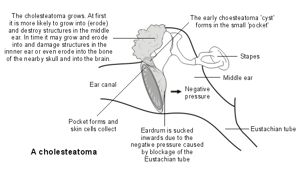
What does a cholesteatoma look like?
In the close-up image below, the collection of small white lumps (white keratin debris) seen on the left-hand side is a cholesteatoma.
ورم كوليسترولي
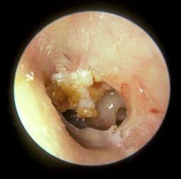
© Michael Hawke MD (Own Work), CC BY 4.0, via Wikimedia Commons
Is a cholesteatoma serious?
If left untreated it will expand further and further inside the ear causing permanent hearing loss, through the inner ear and possibly even next to the brain. Where good medical facilities are available, it would be very unusual for it to get that bad but this can happen in regions with limited healthcare facilities.
Types of cholesteatoma
Congenital cholesteatoma
This is cholesteatoma which is present at the time of birth. For some reason, even though the eardrum is normal, tiny skin cells get sucked into the middle ear, blocking the Eustachian tube.
This then causes long-term fluid in the middle ear (which should usually be free of fluid) and can cause hearing loss. This becomes apparent between the ages of 6 months to 5 years when the child's hearing does not develop properly. This is a very rare condition and the cause is not fully understood.
Acquired cholesteatoma
This type of cholesteatoma develops later, usually in adults between 30 and 50 years old. Again, the cause is not fully understood. Sometimes a cholesteatoma in an adult can arise after having a grommet, a tiny tube that is put through the eardrum as a treatment for middle ear problems, as a child.
Cholesteatoma symptoms
A cholesteatoma grows very gradually, over several months, therefore an early cholesteatoma may have no symptoms.
Other common symptoms include:
Discharge.
Feeling of fullness in the ear.
Discharge
The first symptom is usually a discharge from one ear. It is usually slightly watery, sometimes with a green or yellow colour. The discharge might be slightly smelly and this often looks like an external ear infection (التهاب الأذن الخارجية) or an infection of the inner ear (التهاب الأذن الوسطى) with a perforated eardrum when a doctor examines the ear.
Because it looks just like these common infections, it is usually treated (wrongly) with antibiotic ear drops or pills and although it might get slightly better with these treatments, it never fully clears up. A cholesteatoma usually does not cause pain.
Loss of hearing
After a while, hearing loss can occur in that ear. If the cholesteatoma is left untreated it can spread into the balance organ of the inner ear, causing dizziness or unsteadiness. Ringing in the ear (tinnitus) can also occur.
أعراض أخرى
Eventually, in very rare cases, it can spread right next to the brain and cause an infection of the brain tissue or the lining of the brain. This is very unlikely to occur as most people would seek medical help if they develop the symptoms described above and the cholesteatoma typically grows very slowly.
Causes of a cholesteatoma
We all have skin inside our ear canal. It is meant to be there and is a normal part of our ear. But rarely, the skin right next to the eardrum, deep in the ear, can get sucked in to the middle ear gradually, where it should not be, resulting in a cholesteatoma.
No one quite knows why this happens but it is usually related to the eardrum being drawn inwards, deeper than it is meant to be (retracted).
This skin then forms a tiny pearl, or ball, that keeps burrowing its way deeper into the ear over many months. It damages the delicate bones inside the middle ear - which are responsible for hearing. At this point it becomes painful.
How common is a cholesteatoma?
This condition is uncommon. Around 7-13 people per 100,000 of the population will be diagnosed with a cholesteatoma each year, and it is estimated that an average doctor will see around one new case every 4-5 years.
Cholesteatoma risk factors
Cholesteatoma is more common in men than in women and is usually seen in people who have a history of ear infections. Other risk factors include:
Previous surgery.
Ongoing negative pressure in the middle ear, which may cause the eardrum to become retracted ('pulled in').
Trauma to the ear.
Certain genetic problems such as متلازمة تيرنر أو متلازمة داون.
How is a cholesteatoma diagnosed?
The doctor or ear specialist (ENT doctor) may suspect a cholesteatoma based on the typical symptoms. When the ear is examined with a torch (an otoscope), the cholesteatoma may be seen. Often there is a hole (perforation) in the eardrum (the tympanic membrane) too.
Because the symptoms come on slowly and mimic common ear infections, the diagnosis is often delayed.
It can be very difficult for a doctor to see a cholesteatoma because usually it causes a lot of pus in the ear which blocks the view to the eardrum.
For this reason the diagnosis is usually made by an ear specialist at a hospital.
The ear specialist will use a tiny suction tube to suck away the discharge and look at the eardrum in detail with a microscope that magnifies the view.
By looking in detail at the eardrum, a specialist can usually see the cholesteatoma pushing through the eardrum.
To then see how far it has spread inside the ear, a specialist scan is needed: this is usually a CT scan or an MRI scan.
Do I need any further tests?
Hearing tests (audiometry) may show deafness or hearing loss. These tests are usually performed in a hospital clinic. Samples (swabs) of the ear discharge may also be taken. The discharge often contains a germ (bacterium) called Pseudomonas which is responsible for the smell.
ت فحص الأشعة المقطعية might be needed to see the extent of the damage caused by the cholesteatoma, and to plan further treatment.
Cholesteatoma treatment
Antibiotic eardrops
Antibiotic ear drops can clear away any infection around the cholesteatoma but will not treat the actual problem. Many people will have had antibiotic ear drops prescribed to them without success, before they are diagnosed with a cholesteatoma.
الجراحة
Surgical treatment is carried out by an ear, nose, and throat specialist (an ENT doctor) and this usually consists of an operation under a general anaesthetic. The aim of the surgery is to remove the cholesteatoma and then clear out part of the middle ear so air can circulate around better. This will hopefully stop the cholesteatoma coming back.
There are different types of cholesteatoma surgery that can be done and a specialist ear doctor will advise which operation is best depending on the size of the cholesteatoma and the patient's medical history.
The commonly performed procedures are a mastoidectomy to clear the cholesteatoma from the bone at the back of the ear, the mastoid, or a 'combined approach tympanoplasty' where the damaged part of the eardrum is also removed and replaced..
Other treatment
If the patient is not fit for surgery (for example, if they are very old or frail or have other serious medical conditions) then regular visits to an ear specialist will be recommended to suction out any tiny bits of wax or debris deep in the ear. This will not solve the problem but can keep it from getting worse.
Complications of a cholesteatoma
Untreated, a cholesteatoma will slowly grow and expand. As it grows it can eat into (erode) and destroy anything in its path.
تشمل المضاعفات المحتملة:
Damage and eventual destruction of the tiny bones of the ear (the ossicles). If these are damaged, permanent deafness can occur.
Damage to the mastoid bone. This is the thick bony lump you can feel behind the ear. The mastoid bone is normally filled with pockets of air (a bit like a honeycomb). A cholesteatoma can grow into the mastoid bone, causing infection and destroying it.
Damage to the cochlea and other structures in the inner ear. This can cause permanent deafness on that side, and/or dizziness and balance problems.
Damage to nearby nerves travelling to the face. This can cause weakness (paralysis) of some of the facial muscles.
Cholesteatoma is often infected and this infection can spread to nearby body parts. Very rarely, a cholesteatoma can erode through the skull next to the ear and into the brain. As a result of spreading infection, conditions such as meningitis and brain abscess may develop. These conditions can cause death.
Please note: although a cholesteatoma sounds nasty, it is not cancerous (malignant) and does not spread to distant parts of the body.
ما هي التوقعات؟
This depends on how much damage has been caused by the cholesteatoma by the time it is found and treated. It is also affected by whether any complications such as meningitis or deafness have occurred. The earlier surgery is done, the better the chance of a good outcome.
If you have had a cholesteatoma, you will need to be followed up in an ENT clinic.
Can a cholesteatoma come back?
If the ear starts discharging again, further surgery may be required. MRI scans are increasingly being used, rather than surgery to review and see whether a cholesteatoma has formed again.
A cholesteatoma grows very gradually, over several months.
How do we hear?
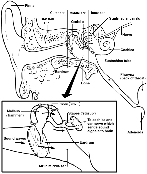
The ear is divided into three parts - the external ear, the middle ear and the inner ear. The middle ear, which is behind the eardrum (the tympanic membrane) is filled with air. Air comes from the back of the nose up a thin channel called the Eustachian tube.
In the middle ear there are three tiny bones (ossicles) - the hammer (malleus), anvil (incus) and stirrup (stapes). The inner ear includes the cochlea and the balance organ which contains the semicircular canals, the utricle and saccule.
Sound waves come into the external ear and hit the eardrum. The sound waves cause the eardrum to vibrate. The sound vibrations pass from the eardrum to the ossicles. The ossicles then transmit the vibrations to the cochlea in the inner ear.
The cochlea converts the vibrations to sound signals which are sent along a nerve from the ear to the brain, allowing us to hear.
The semicircular canals and two additional structures called the utricle and saccule in the inner ear all contain a fluid that moves around as we move into different positions. The movement of the fluid is sensed by tiny hairs in the semicircular canals and the utricle and saccule which send messages to the brain along the ear nerve to help maintain balance and posture.
Detail of middle ear
اختيارات المرضى لـ مشاكل السمع

الأذن والأنف والحنجرة
فقدان السمع لدى كبار السن
معظم الأشخاص الذين تزيد أعمارهم عن 60 عامًا يعانون من فقدان السمع بدرجة معينة. يمكن أن تختلف شدة الفقدان بشكل كبير من شخص لآخر. يمكن لأجهزة السمع والمعدات المساعدة عند فقدان السمع أن تحدث فرقًا كبيرًا في جودة حياتك. إذا كنت قلقًا بشأن سمعك، فلا تعاني في صمت - ناقش هذا مع طبيبك.
بقلم الدكتورة ماري هاردينغ، MRCGP

الأذن والأنف والحنجرة
شمع الأذن
A build-up of earwax can cause reduced hearing and sometimes other symptoms. Earwax can usually be easily removed.
بقلم الدكتورة فيليبا فينسنت، MRCGP
الأسئلة الشائعة
ما هي فترة التعافي النموذجية بعد جراحة الكوليستياتوما؟
تذكر المقالة أن الجراحة تهدف إلى إزالة الكوليستياتوما وتنظيف جزء من الأذن الوسطى لمنع عودتها. كما تشير إلى أنك ستحتاج إلى مواعيد متابعة في عيادة الأنف والأذن والحنجرة. بعد الجراحة، يعتمد التوقع على مدى الضرر قبل العلاج.
هل هناك أي تغييرات معينة في نمط الحياة يجب أن أقوم بها إذا كان لدي كوليستياتوما؟
لا يحدد المقال أي تغييرات معينة في نمط الحياة مطلوبة لعلاج الكوليستياتوما. يركز بشكل أساسي على التشخيص، العلاج من خلال الجراحة، وإدارة المضاعفات. غالبًا ما يرتبط هذا المرض بتغيرات الضغط المزمنة في الأذن أو يمكن أن يكون خلقيًا.
كم من الوقت يستغرق ظهور أعراض ملحوظة للكوليستياتوما؟
ينمو الورم الكوليسترولي بشكل تدريجي جداً على مدى عدة أشهر، مما يعني أن الورم المبكر قد لا يكون له أي أعراض. يمكن أن تظل الحالة بدون أعراض لفترة من الوقت قبل أن تتطور إلى إفرازات، فقدان السمع، أو دوار.
إذا أجريت لي جراحة الكولستيتيتوما، كم مرة سأحتاج إلى مواعيد متابعة؟
إذا كنت قد أصبت بورم كوليسترولي، فستحتاج إلى متابعة في عيادة الأنف والأذن والحنجرة. لم يتم تحديد تكرار هذه المتابعات في المقالة، لكنها مهمة لمراقبة ما إذا كان الورم الكوليسترولي قد يتشكل مرة أخرى.
هل يمكن للأطفال الإصابة بالكوليستياتوما؟
نعم، يمكن للأطفال أن يصابوا بالكوليستياتوما. الكوليستياتوما الخلقية تكون موجودة عند الولادة وتظهر بين 6 أشهر و5 سنوات إذا لم يتطور سمع الطفل بشكل صحيح. هذا النوع نادر جدًا وسببه غير مفهوم بالكامل.
قراءة إضافية ومراجع
- ورم كوليسترولي; NICE CKS، أغسطس 2024 (الوصول متاح فقط في المملكة المتحدة)
- Castle JT; Cholesteatoma Pearls: Practical Points and Update. Head Neck Pathol. 2018 Sep;12(3):419-429. doi: 10.1007/s12105-018-0915-5. Epub 2018 Aug 1.
- Ayache D, Darrouzet V, Dubrulle F, et al; Imaging of non-operated cholesteatoma: clinical practice guidelines. Eur Ann Otorhinolaryngol Head Neck Dis. 2012 Jun;129(3):148-52. doi: 10.1016/j.anorl.2011.09.005. Epub 2012 Feb 7.
عن المؤلفعرض السيرة الذاتية الكاملة

الدكتور دوغ مكيتشني، MRCGP
كاتب طبي
MA, MBBS, MSc, DRCOG, MRCP(UK), MRCGP(2021), FHEA
الدكتور دوغ مكيتشني هو طبيب عام في هيئة الخدمات الصحية الوطنية يعمل في لندن. يعمل بدوام كامل في المجال السريري وهو أيضًا نائب رئيس وحدة الممارسة السريرية والمهنية في كلية الطب بجامعة كوليدج لندن.
حول المراجععرض السيرة الذاتية الكاملة

الدكتور كولين تايدي، MRCGP
طبيب عام، مؤلف طبي
MBBS, MRCGP, MRCP (Paediatrics), DCH
الدكتور كولين تايدي هو طبيب في هيئة الخدمات الصحية الوطنية، ويعمل في أوكسفوردشاير.
تاريخ المقال
تمت كتابة المعلومات على هذه الصفحة ومراجعتها من قبل أطباء مؤهلين.
المقال متاح أيضًا باللغة الإنجليزية, الألمانية, إسبانية, الفرنسية, إيطالي, البرتغالية, الهندية, العبرية, العربية ,، و السويدية.
المراجعة التالية مستحقة: 2 فبراير 2028
3 فبراير 2025 | أحدث إصدار

اسأل، شارك، تواصل.
تصفح المناقشات، اطرح الأسئلة، وشارك التجارب عبر مئات المواضيع الصحية.

هل تشعر بتوعك؟
قم بتقييم أعراضك عبر الإنترنت مجانًا
اشترك في النشرة الإخبارية للمرضى
جرعتك الأسبوعية من النصائح الصحية الواضحة والموثوقة - مكتوبة لمساعدتك على الشعور بالاطلاع والثقة والتحكم.
من خلال الاشتراك، فإنك تقبل سياسة الخصوصية. يمكنك إلغاء الاشتراك في أي وقت. نحن لا نبيع بياناتك أبدًا.