فحص النظائر المشعة
Isotope scan
مراجعة من قبل الدكتور كريشنا فاخاريا، MRCGPآخر تحديث بواسطة الدكتورة راشيل هدسون، MRCGPآخر تحديث 24 أغسطس 2023
يتوافق مع الإرشادات التحريرية
- تنزيلتنزيل
- مشاركة
- Language
- نقاش
- Ask Patient
- نسخة صوتية
- أضف إلى المصادر المفضلة على جوجل
A radionuclide scan is a way of imaging bones, organs and other parts of the body by using a small dose of a radioactive chemical. There are different types of radionuclide chemical. The one used depends on which organ or part of the body is to be scanned.
ملاحظة: المعلومات أدناه هي دليل عام فقط. قد تختلف الترتيبات وطريقة إجراء الاختبارات بين المستشفيات المختلفة. دائماً اتبع التعليمات المقدمة من طبيبك أو المستشفى المحلي.
نظرة سريعة
A radionuclide scan uses a small amount of a radioactive chemical to create images of inside your body.
It can help detect conditions like cancer, infection, blood clots, or assess organ function.
The chemical usually enters your body by injection or by swallowing, and a special camera detects its signals.
Preparation may involve drinking fluids or pausing certain medications.
The amount of radiation used is small and generally considered safe.
Tell your doctor if you are pregnant or might be, as pregnant women should not have these scans.
Why do you have a radionuclide scan?
A radionuclide scan may be done for all sorts of reasons. For example:
A bone scan is a common type. A radionuclide is used which collects in areas where there is a lot of bone activity (where bone cells are breaking down or repairing parts of the bone). So a bone scan is used to detect areas of bone where there is cancer, infection, or damage. These areas of activity are seen as 'hot spots' on the scan picture. See the separate leaflet called Bone Scan for more details.
A kidney scan can assess how well a kidney is working (as the radionuclide chosen is taken up by kidney cells and passes into the urine). So, the scan can detect scars on the kidney and how well urine drains from the kidney to the bladder. See the separate leaflet called DMSA Scan for more details.
Lung perfusion scan (also called a 'VQ scan') can detect blood clots in the lungs (pulmonary embolism).
A heart scan can assess blood flow to the heart muscle. Areas of poor blood flow to the heart muscle do not 'take up' the radionuclide very well and this will be shown in the picture.
A thyroid scan may be done to assess cases of overactive thyroid (hyperthyroidism). For example, some nodules (small 'lumps') are sometimes a focus of overactivity and will show as 'hot spots' on the picture. See the separate leaflet called Thyroid Scans and Uptake Tests for more details.
Lacrimal scintigraphy is done to test the function of tear ducts (lacrimal ducts.) The radionuclide is given as eye drops.
Lymphoscintigraphy is done to check the drainage of the العقد الليمفاوية in people with a type of swelling of the legs, called الوذمة اللمفية.
There are various other types of radionuclide tests.
What preparation do I need?
The preparation needed is usually very little. It will depend on which type of scan you are having. Your local hospital should give you specific information to help you prepare for these tests.
For some types of scan, you may be asked to have lots to drink to help to flush the radionuclide through your body.
For some types of scan you may also be asked to empty your bladder of urine before the scanning begins.
For some scans, such as thyroid scans, you may be instructed to stop certain medications for some time before the scan.
As these tests involve a small amount of radiation, pregnant women should not have them.
ملاحظة: let your doctor know if you are, or think you could be, pregnant. You should also let your doctor know if you are breastfeeding.
What happens during a radionuclide scan?
The procedures for the different types of radionuclide scans are different. Information about your scan should be sent to you with the appointment.
Depending on the type of scan you have, you usually either swallow a small quantity of radionuclide, or it is injected into a vein in your arm.
It then takes some time - sometimes several hours (depending on what is being scanned) - for the radionuclide to travel to the target organ or tissue, and to be 'taken' into the active cells.
So, after receiving the radionuclide you may have a wait of a few hours. You may be able to go out and come back to the scanning room later in the day.
When it is time to do the scanning, you usually lie on a couch while the gamma camera detects the gamma rays coming from your body.
Gamma camera
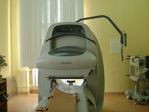
The computer turns the information into a picture. You need to lie as still as possible whilst each picture is taken (so it is not blurred). Some pictures can take 20 minutes or more to expose.
The number of pictures taken and the time interval between each picture vary depending on what is being scanned. Sometimes only one picture is needed. However, for some scans (such as bone scans or heart scans), two or more pictures are needed. Each picture may be taken several hours apart. So, the whole process can take several hours.
What happens after a radionuclide scan?
Radionuclide scans do not generally cause any side-effects.
Uncommon side-effects from radionuclides may include flushing, racing heart and الغثيان but these are short-lived because they are flushed out of your system quickly.
Through the natural process of radioactive decay, the small amount of radioactive chemical in your body will lose its radioactivity over time. It may also pass out of your body through your urine or poo during the first few hours or days following the test.
You may be instructed to take special precautions after urinating, to flush the toilet twice and to wash your hands thoroughly. You may be advised to drink plenty of water to help flush the chemicals out of your system.
إذا كنت على اتصال مع الأطفال أو النساء الحوامل، يجب أن تخبر طبيبك. على الرغم من أن مستويات الإشعاع المستخدمة في الفحص صغيرة، قد ينصحون باتخاذ احتياطات خاصة. يجب أن يقدم لك المستشفى المزيد من النصائح حول هذا الأمر.
Are there risks with radionuclide scans?
The term 'radioactivity' may sound alarming. But, the radioactive chemicals used in radionuclide scans are considered to be safe, and they leave the body quickly in the urine. The dose of radiation that your body receives is very small.
In many cases, the level of radiation involved is not much different to a series of a few normal X-rays. However:
كما هو الحال مع أي نوع آخر من الإشعاع (مثل الأشعة السينية), there is a small risk that the gamma rays may affect an unborn child. So, tell your doctor if you are pregnant or if you may be pregnant.
Rarely, some people have an allergic reaction to the injected chemical. Tell your doctor if you are allergic to iodine.
Theoretically, it is possible to receive an overdose when the chemical is injected. This is very rare.
How does a radionuclide scan work?
A radionuclide (sometimes called a radioisotope or isotope) is a chemical which emits a type of radioactivity called gamma rays. A tiny amount of radionuclide is put into the body, usually by an injection into a vein. Sometimes it is breathed in, or swallowed, or given as eye drops, depending on the test.
There are different types of radionuclides. Different ones tend to collect or concentrate in different organs or tissues. So, the radionuclide used depends on which part of the body is to be scanned.
For example, if radioactive iodine is injected into a vein it is quickly taken up into the tissues of the thyroid gland. So, it is used to scan the thyroid gland.
Cells which are most 'active' in the target tissue or organ will take up more of the radionuclide. So, active parts of the tissue will emit more gamma rays than less active or inactive parts.
Gamma rays are similar to X-rays and are detected by a device called a gamma camera. The gamma rays which are emitted from inside the body are detected by the gamma camera, are converted into an electrical signal and sent to a computer.
The computer builds a picture by converting the differing intensities of radioactivity emitted into different colours or shades of grey. This is seen below in a lung perfusion scan.
Lung perfusion scan
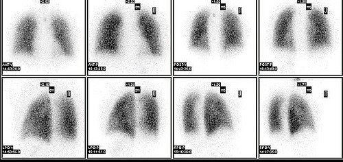
Alternatively areas of the target organ or tissue which emit lots of gamma rays may be shown as red spots ('hot spots') on the picture on the computer monitor.
Areas which emit low levels of gamma rays may be shown as blue ('cold spots'). Various other colours may be used for 'in between' levels of gamma rays emitted.
اختيارات المرضى لـ التصوير
الفحوصات والتحقيقات
فحص العظام
A bone scan is a special type of nuclear medicine procedure that uses radionuclides to create a picture of the bones. Radionuclides are chemicals which emit radioactivity that can be detected by special scanners. A bone scan is different to a bone density scan (DEXA). See the DEXA Scan leaflet for more information on this bone density test. A DEXA scan is also known as a DXA scan. Note: the information below is a general guide only. The arrangements, and the way tests are performed, may vary between different hospitals. Always follow the instructions given by your doctor or local hospital.
بقلم الدكتور دوغ مكيتشني، MRCGP
الفحوصات والتحقيقات
فحص الأشعة المقطعية
Note: the information below is a general guide only. The arrangements, and the way tests are performed, may vary between different hospitals. Always follow the instructions given by your doctor or local hospital.
بقلم الدكتورة راشيل هدسون، MRCGP
الأسئلة الشائعة
What is a radionuclide?
A radionuclide, also known as a radioisotope or isotope, is a chemical that gives off gamma rays. A small amount of this chemical is introduced into the body to allow a gamma camera to create images of internal organs or tissues. Different radionuclides are used depending on the specific organ or tissue being scanned.
Are there specific radionuclides for different body parts?
Yes, there are various types of radionuclides, and each tends to gather in specific organs or tissues. The choice of radionuclide depends on which part of the body needs to be scanned. For instance, radioactive iodine is used for thyroid gland scans because it's readily absorbed by thyroid tissues.
How are the images from a radionuclide scan interpreted?
The gamma camera detects the gamma rays emitted from inside your body and converts them into electrical signals for a computer. The computer then creates a picture, using different colours or shades of grey to represent varying intensities of radioactivity. "Hot spots" (often shown in red) indicate areas with high gamma ray emission, meaning high activity, while "cold spots" (often in blue) indicate low emission or activity. Other colours might be used for intermediate levels.
Can I have a radionuclide scan if I am breastfeeding?
If you are breastfeeding, you should inform your doctor before having a radionuclide scan. They will be able to advise you on any special precautions that may be necessary.
What happens if I have an allergic reaction to the chemical injected during the scan?
Allergic reactions to the injected chemical are rare. However, if you have any known allergies, especially to iodine, you should inform your doctor beforehand.
Will I experience any discomfort during the scan procedure itself?
During the scanning itself, you usually lie still on a couch while the gamma camera takes pictures. You'll need to remain as still as possible to prevent blurring of the images. Some pictures can take 20 minutes or longer to expose.
قراءة إضافية ومراجع
- Myocardial perfusion scintigraphy for the diagnosis and management of angina and myocardial infarction; NICE Technology Appraisal Guidance, November 2003 (last updated July 2011)
- Nuclear Medicine; Radiology.info.org (American College of Radiology and Radiological Society of North America)
- Guidelines; British Nuclear Medicine Society
- Gruning T, Drake BE, Freeman SJ; Single-photon emission CT using (99m)Tc-dimercaptosuccinic acid (DMSA) for characterization of suspected renal masses. Br J Radiol. 2014 Jul;87(1039):20130547. doi: 10.1259/bjr.20130547. Epub 2014 May 16.
- Radionuclide bone scans/bone scintigraphy; أبحاث السرطان في المملكة المتحدة
- Myocardial perfusion scans; مؤسسة القلب البريطانية
عن المؤلفعرض السيرة الذاتية الكاملة
تم التحديث بواسطة

الدكتورة راشيل هدسون، MRCGP
طبيب عام وكاتب طبي
بكالوريوس الطب والجراحة، زميل الكلية الملكية للممارسين العامين (2008)، بكالوريوس العلوم (العلوم الطبية)، دبلوم في الصحة الجنسية والإنجابية، دبلوم الكلية الملكية لأطباء النساء والتوليد، دبلوم صحة الطفل
الدكتورة راشيل هدسون، طبيبة عامة في هيئة الخدمات الصحية الوطنية تعمل في شمال غرب إنجلترا.
حول المراجععرض السيرة الذاتية الكاملة

الدكتور كريشنا فاخاريا، MRCGP
كبير المسؤولين الطبيين للصحة، أوبتوم المملكة المتحدة
MBChB, MRCGP(2013), BMedSci (hons), DFSRH, DRCOG, PGDipDerm (Distn)
الدكتورة كريشنا فاخاريا هي طبيبة عامة في هيئة الخدمات الصحية الوطنية. كما أنها ممتحنة منتظمة لدبلوم الدراسات العليا في الأمراض الجلدية العملية في جامعة كارديف، بالإضافة إلى كونها كبيرة المسؤولين الطبيين للصحة في Optum UK.
تاريخ المقال
المقال متاح أيضًا باللغة الإنجليزية, الألمانية, إسبانية, الفرنسية, إيطالي, البرتغالية, الهندية, العبرية, العربية ,، و السويدية.
موعد المراجعة التالية: 22 أغسطس 2028
24 أغسطس 2023 | أحدث إصدار
شارك أفكارك أو تجاربك مع الآخرين في مجتمعات المرضى.
وصول بسيط إلى العلاجات والخدمات الصحية الموثوقة عبر الإنترنت.
Common conditions
Get help with common conditions through the NHS, without a GP appointment.
 التهاب الجيوب الأنفية الحاد
 التهاب اللوزتين البكتيري
 القوباء
 لدغات الحشرات
 الحزام الناريعدوى المسالك البولية
اشترك في النشرة الإخبارية للمرضى
جرعتك الأسبوعية من النصائح الصحية الواضحة والموثوقة - مكتوبة لمساعدتك على الشعور بالاطلاع والثقة والتحكم.
من خلال الاشتراك، فإنك تقبل سياسة الخصوصية. يمكنك إلغاء الاشتراك في أي وقت. نحن لا نبيع بياناتك أبدًا.