تنظير المعدة
تنظير داخلي
مراجعة من قبل الدكتورة هايلي ويلاسي، زميلة الكلية الملكية للأطباء العامين آخر تحديث بواسطة الدكتورة فيليبا فينسنت، MRCGPآخر تحديث 3 يوليو 2024
يتوافق مع الإرشادات التحريرية
- تنزيلتنزيل
- مشاركة
- Language
- نقاش
- نسخة صوتية
- أضف إلى المصادر المفضلة على جوجل
Gastroscopy is a test to look inside the gullet (oesophagus), the stomach and the first part of the gut (small intestine) known as the duodenum.
ملاحظة: the information below is a general guide only. The arrangements, and the way tests are performed, may vary between different hospitals and areas. Always follow the instructions given by your doctor or local hospital.
نظرة سريعة
التنظير المعدي هو اختبار لفحص داخل المريء والمعدة والجزء الأول من الأمعاء الدقيقة.
يتم تمرير أنبوب رفيع ومرن مزود بكاميرا عبر فمك وصولاً إلى معدتك.
يمكن أن يساعد في تحديد أسباب عسر الهضم، وحرقة المعدة، وآلام البطن، أو صعوبة البلع.
عادةً ما تستغرق العملية حوالي 10 دقائق، ولكن يُفضل تخصيص ساعتين على الأقل لموعدك.
سيُطلب منك عدم الأكل أو الشرب لمدة 4-6 ساعات قبل الاختبار.
اطلب المساعدة الطبية الفورية إذا كنت تعاني من تفاقم ألم البطن، أو حمى، أو صعوبة في التنفس، أو تقيؤ دم بعد الاختبار.
What is a gastroscopy?
تنظير المعدة
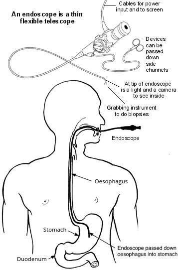
A gastroscopy is a test where an operator - usually a doctor or nurse - looks into the upper part of the gut (the upper gastrointestinal tract) with a camera.
The upper gut consists of the gullet (oesophagus), the stomach and the first part of the gut (small intestine) known as the duodenum. The operator uses an endoscope to look inside the gut.
An endoscope is a thin, flexible telescope. It is about as thick as a little finger. The endoscope is passed through the mouth, down the food pipe into the oesophagus and down towards the stomach and duodenum.
The tip of the endoscope contains a light and a tiny video camera so the operator can see inside the gut.
The endoscope also has a side channel down which various instruments can pass. These can be manipulated by the operator. For example, the operator may take a small sample (خزعة) from the inside lining of the stomach by using a thin 'grabbing' instrument which is passed down a side channel.
The procedure is sometimes called an "upper GI endoscopy" - this refers to the upper gastro-intestinal tract.
Why do I need a gastroscopy?
A gastroscopy may be advised for symptoms such as:
Repeated (recurring) indigestion.
Recurring heartburn.
Pains in the upper tummy (abdomen).
Repeatedly being sick (vomiting).
صعوبة في البلع.
Other symptoms thought to be coming from the upper gut, stomach or oesophagus.
What is a gastroscopy looking for?
Inflammation of the gullet (oesophagus), called oesophagitis. The operator will see areas of redness on the lining of the oesophagus.
قرحة المعدة و قرحة الاثني عشر. An ulcer looks like a small, red crater on the inside lining of the stomach or on the first part of the gut (small intestine) known as the duodenum.
Inflammation of the duodenum (duodenitis) and inflammation of the stomach (التهاب المعدة).
Stomach و oesophageal cancer.
فتق الحجاب الحاجز
Various other rare conditions.
What happens during a gastroscopy?
Gastroscopy is usually done as an outpatient 'day case'. It is a routine test which is commonly done. Before the test the operator will explain what is going to happen and ask for a consent form to be signed.
The operator may numb the back of the throat by spraying on some local anaesthetic or giving an anaesthetic lozenge to suck. A sedative may also be given to help relaxation. This is usually given by an injection into a vein in the back of the hand. The sedative causes drowsiness but is not a general anaesthetic and does not put someone to sleep.
You lie on your side on a couch. You are asked to put a plastic mouth guard between your teeth. This works to protect your teeth and stops you biting the endoscope. The operator will then ask you to swallow the first section of the endoscope. Modern endoscopes are quite thin but some people may find this difficult.
The operator then gently pushes it further down the gullet (oesophagus) and into the stomach and the first part of the gut (small intestine) known as the duodenum. The video camera at the tip of the endoscope sends pictures to a screen.
The operator watches the screen for abnormalities of the oesophagus, stomach and duodenum. Air is passed down a channel in the endoscope into the stomach to make the stomach lining easier to see. This may cause a bloating sensation and belching.
The operator may take one or more small tissue samples (biopsies) of parts of the inside lining of the gut - depending on why the test is done and what they see on the screen. This is painless. The biopsy samples are sent to the laboratory for testing and to look at under the microscope. The endoscope is then gently pulled out.
How long does a gastroscopy take?
A gastroscopy usually takes about 10 minutes. However, at least two hours should be allowed for the whole appointment. This is to prepare, give time for the sedative to work, for the gastroscopy itself and to recover. A gastroscopy may be quite uncomfortable but it does not usually hurt.
How do you prepare for a gastroscopy?
You should receive instructions from the hospital department before your test. The sort of instructions given commonly include:
Avoid eating or drinking for 4-6 hours before the test. The stomach needs to be empty. (Small sips of water may be allowed up to two hours before the test.)
If you have a sedative you will need somebody to accompany you home.
Advice about medication which may need to be stopped before the test.
What can I expect after a gastroscopy?
Most people are ready to go home after resting for half an hour or so.
If you have had a sedative - you may take a bit longer to be ready to go home. The sedative will normally make you feel quite pleasant and relaxed. However, you should not drive, operate machinery or drink alcohol for 24 hours after having the sedative. You will need somebody to accompany you home and to stay with you for 24 hours until the effects have fully worn off. Most people are able to resume normal activities after 24 hours.
The operator writes a report and sends it to the doctor who requested the gastroscopy, usually a gastroenterologist but occasionally another specialist or GP. The result from any sample (biopsy) may take some days or even weeks, which can delay the report being sent. The operator may inform you about what they saw before you leave. However, if you have had a sedative you may not remember afterwards what you were told. Therefore, you may wish to have a relative or close friend with you who may be able to help remember what was said.
Is gastroscopy reliable?
Gastroscopy is a good test for seeing abnormalities in the upper gut. However, it is not fool-proof. For example, gastroscopy may not detect a small number of cases of early ulcers or early cancer. Sometimes a repeat gastroscopy may be advised if symptoms persist or become worse, even if a previous gastroscopy was reported as normal.
Is a gastroscopy dangerous?
Most gastroscopies are done without any problem. Some people have a mildly sore throat for a day or so afterwards. You may feel tired or sleepy for several hours if you have a sedative. There is a slight risk of developing a chest infection or pneumonia following a gastroscopy.
Occasionally, the endoscope causes some damage to the gut. This may cause bleeding, infection and rarely a hole (perforation). If any of the following occur within 48 hours after a gastroscopy, seek immediate medical help:
ألم البطن (المعدة). (In particular, if it becomes gradually worse, and is different from or more intense than any 'usual' indigestion pains or heartburn.)
Raised temperature (الحمى).
صعوبة في التنفس.
Vomiting blood.
A small number of people have a نوبة قلبية أو السكتة الدماغية during, or soon after, a gastroscopy. These tend to be older people who are already in poor health. These serious complications are rare in most people who are otherwise reasonably healthy.
Rarely, some people have an allergic reaction to the sedation.
اختيارات المرضى لـ تنظير داخلي

الجراحة والإجراءات
تنظير السيني
A sigmoidoscopy is a test that looks at the rectum and lower part of the large intestine. Note: the information below is a general guide only. The arrangements and the way tests are performed, may vary between different hospitals. Always follow the instructions given by your doctor or local hospital.
بقلم الدكتورة كلوديا بيرتي، MRCGP

الجراحة والإجراءات
تنظير القصبات
تنظير القصبات هو اختبار يمكن أن يساعد في تشخيص وعلاج حالات التنفس لديك. يتضمن ذلك إدخال الطبيب لأنبوب كاميرا مرن إلى رئتيك. ملاحظة: المعلومات أدناه هي دليل عام فقط. الترتيبات وطريقة إجراء الاختبارات تختلف غالبًا بين المستشفيات المختلفة.
بقلم الدكتورة توني هازيل، FRCGP
الأسئلة الشائعة
ما هو الغرض من واقي الفم البلاستيكي المستخدم أثناء تنظير المعدة؟
أثناء إجراء تنظير المعدة، يتم وضع واقي فم بلاستيكي بين أسنانك. الغرض منه هو حماية أسنانك ومنعك من عض المنظار عن طريق الخطأ أثناء الإجراء.
كيف يرى الطبيب داخل الأمعاء أثناء تنظير المعدة؟
المنظار، وهو أنبوب رفيع ومرن، يحتوي على كاميرا فيديو صغيرة وضوء في طرفه. ترسل هذه الكاميرا صورًا حية إلى شاشة، مما يسمح للمشغل برؤية البطانة الداخلية لمريئك ومعدتك والجزء الأول من الأمعاء الدقيقة.
ماذا يحدث لعينات الأنسجة (الخزعات) التي تؤخذ أثناء تنظير المعدة؟
إذا تم أخذ عينات صغيرة من الأنسجة (خزعات) أثناء التنظير المعدي، يتم إرسالها إلى مختبر للفحص. هناك، يتم فحصها تحت المجهر للمساعدة في تشخيص أي شذوذ.
لماذا قد أشعر بالانتفاخ أو التجشؤ أثناء تنظير المعدة؟
أثناء الإجراء، يتم تمرير الهواء عبر قناة في المنظار إلى معدتك. يتم ذلك للمساعدة في جعل بطانة المعدة أسهل للرؤية من قبل المشغل، ولكن قد يسبب لك شعورًا بالانتفاخ ويؤدي إلى التجشؤ.
هل يمكنني الحصول على نتائج تنظير المعدة فور الانتهاء من الإجراء؟
قد يقوم المشغل بإبلاغك بنتائجهم فورًا بعد الإجراء. ومع ذلك، إذا كنت قد تلقيت مهدئًا، فقد لا تتذكر ما تمت مناقشته. أيضًا، إذا تم أخذ أي عينات نسيجية (خزعات)، فقد يتأخر التقرير الكامل حيث قد يستغرق الأمر عدة أيام أو حتى أسابيع لعودة تلك النتائج من المختبر.
إذا كانت نتائج تنظير المعدة طبيعية، هل يعني ذلك بالتأكيد أنني لا أعاني من مشكلة؟
على الرغم من أن تنظير المعدة يعتبر موثوقًا بشكل عام، إلا أنه ليس دائمًا دقيقًا تمامًا. في عدد قليل من الحالات، قد لا يكتشف القرح المبكرة جدًا أو السرطان في مراحله المبكرة. إذا استمرت الأعراض لديك أو تفاقمت، حتى بعد إجراء تنظير معدة طبيعي، قد يوصي طبيبك بإجراء الفحص مرة أخرى.
قراءة إضافية ومراجع
عن المؤلفعرض السيرة الذاتية الكاملة

الدكتورة فيليبا فينسنت، MRCGP
طبيب عام، مؤلف طبي
MB BS, Bsc, MRCGP (2000), DCH, DFSRH, DRCOG
الدكتورة فيليبا فينسنت هو طبيب عام في هيئة الخدمات الصحية الوطنية يعمل في شمال لندن.
حول المراجععرض السيرة الذاتية الكاملة

الدكتورة هايلي ويلاسي، زميلة الكلية الملكية للأطباء العامين
طبيب عام، مؤلف طبي
MBChB (1992), DRCOG, DFFP, MRCOG (Part 1) MRCGP (2007), DFSRH (2013), MSc - medical education (2020)
كانت الدكتورة هايلي ويلاسي طبيبة عامة في هيئة الخدمات الصحية الوطنية تعمل في شمال غرب إنجلترا، وتقاعدت من الممارسة السريرية في عام 2022 بعد 30 عامًا.
تاريخ المقال
تمت كتابة المعلومات على هذه الصفحة ومراجعتها من قبل أطباء مؤهلين.
المقال متاح أيضًا باللغة الإنجليزية, الألمانية, إسبانية, الفرنسية, إيطالي, البرتغالية, الهندية, العبرية, العربية ,، و السويدية.
المراجعة التالية مستحقة: 2 يوليو 2027
3 يوليو 2024 | أحدث إصدار
آخر تحديث بواسطة
الدكتورة فيليبا فينسنت، MRCGPمراجعة من قبل
الدكتورة هايلي ويلاسي، زميلة الكلية الملكية للأطباء العامين

اسأل، شارك، تواصل.
تصفح المناقشات، اطرح الأسئلة، وشارك التجارب عبر مئات المواضيع الصحية.

هل تشعر بتوعك؟
قم بتقييم أعراضك عبر الإنترنت مجانًا
اشترك في النشرة الإخبارية للمرضى
جرعتك الأسبوعية من النصائح الصحية الواضحة والموثوقة - مكتوبة لمساعدتك على الشعور بالاطلاع والثقة والتحكم.
من خلال الاشتراك، فإنك تقبل سياسة الخصوصية. يمكنك إلغاء الاشتراك في أي وقت. نحن لا نبيع بياناتك أبدًا.
المزيد في الجراحة والإجراءات
- التخدير
- التخدير لاستبدال الورك أو الركبة
- البوتوكس
- تنظير المهبل وعلاجات عنق الرحم
- جراحة تحويل مسار الشريان التاجي
- الموت أو تلف الدماغ بسبب التخدير
- تجلط الأوردة العميقة
- خزعة بطانة الرحم
- تخفيف الألم باستخدام الإيبيدورال بعد الجراحة
- تنظير البطن والجراحة بالمنظار
- جراحة العيون بالليزر
- تعزيز الشفاه
- التخدير الموضعي لعملية العين الخاصة بك
- تلف الأعصاب بعد حقن فوق الجافية
- جراحة تصغير الأنف (تجميل الأنف)
- الانسداد الرئوي
- تنظير السيني
- التخدير النخاعي
- الجراحة الصدرية بمساعدة الفيديو
- جراحة فقدان الوزن