الانصباب الجنبي
مراجعة من قبل الدكتور دوغ مكيتشني، MRCGPآخر تحديث بواسطة الدكتورة هايلي ويلاسي، زميلة الكلية الملكية للأطباء العامين آخر تحديث 21 ديسمبر 2023
يتوافق مع الإرشادات التحريرية
- تنزيلتنزيل
- مشاركة
- Language
- نقاش
- نسخة صوتية
- أضف إلى المصادر المفضلة على جوجل
الانصباب الجنبي هو تجمع للسوائل بجانب الرئة. هناك أسباب مختلفة لذلك. قد يسبب الانصباب ضيقًا في التنفس. يمكن تصريف السوائل إذا لزم الأمر. يهدف العلاج بشكل رئيسي إلى معالجة السبب الأساسي.
نظرة سريعة
الانصباب الجنبي هو تراكم السوائل في المساحة بين الرئة وجدار الصدر.
يمكن أن يكون سبب هذه الحالة عوامل مختلفة، بما في ذلك العدوى، وفشل القلب، وبعض أنواع السرطان.
يمكن أن تشمل الأعراض ألمًا في الصدر وضيقًا في التنفس، خاصة مع زيادة السوائل.
عادةً ما يؤكد تصوير الصدر بالأشعة السينية وجود انصباب جنبي.
غالبًا ما يركز العلاج على السبب الأساسي، ولكن يمكن أيضًا تصريف السائل نفسه.
يمكن أن تساعد الإجراءات مثل التصاق الجنبة أو المصارف الدائمة في إدارة الانصبابات المتكررة.
ما هو الانصباب الجنبي؟
الرئتان والمجاري الهوائية مع الغشاء البلوري
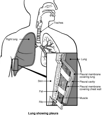
الانصباب الجنبي يعني تراكم السوائل في المساحة بين الرئة وجدار الصدر.
الغشاء البلوري هو غشاء رقيق يبطن الجزء الداخلي من جدار الصدر ويغطي الرئتين. عادة ما يكون هناك كمية صغيرة من السائل بين طبقتي الغشاء البلوري. يعمل هذا السائل مثل الزيت المزلّق بين الرئتين وجدار الصدر أثناء حركتهما عند التنفس.
يتطور الانصباب الجنبي عندما يتراكم هذا السائل ويفصل الرئة عن جدار الصدر.
أنواع الانصباب الجنبي
يتم تصنيف الانصباب الجنبي إلى ترشحات أو رشحات بناءً على كمية البروتين التي تحتويها.
التسربات تحتوي على مستوى منخفض من البروتين أقل من <25 جم/لتر. يتراكم السائل بسبب اضطراب في توازن ضغوط السوائل.
الإفرازات تحتوي على مستوى عالٍ من البروتين يتجاوز 35 جم/لتر. يتراكم السائل بسبب زيادة تسرب (نفاذية) الأوعية الدموية الصغيرة.
الانصباب الجنبي النتحي
غالبًا ما تحدث هذه بسبب حالات مثل فشل القلب وفشل الكبد. الأسباب الأقل شيوعًا هي متلازمة الكلوية, قصور الغدة الدرقية وكذلك نتيجة لغسيل الكلى البريتوني.
الانصباب الجنبي النضحي
الأسباب الأكثر شيوعًا لهذه هي العدوى، مثل الالتهاب الرئوي أو السل ,، أو السرطان. تشمل الأسباب الأقل شيوعًا الحالات المناعية الذاتية مثل التهاب المفاصل الروماتويدي ,، بعد نوبة قلبية و التهاب البنكرياس.
أسباب الانصباب الجنبي
الانصباب الجنبي هو مضاعفة لحالات مختلفة. فيما يلي بعض الأسباب الأكثر شيوعًا للانصباب الجنبي (ولكن هناك أسباب نادرة أخرى أيضًا):
عدوى الرئة (الالتهاب الرئوي), السل ,، وقد تسبب السرطانات التهاب الرئة والجنبة. قد يؤدي ذلك إلى تراكم السوائل في انصباب جنبي.
بعض حالات التهاب المفاصل قد تسبب التهاب الغشاء البلوري بالإضافة إلى التهاب المفاصل. على سبيل المثال، الانصباب الجنبي هو مضاعفة غير شائعة لـ التهاب المفاصل الروماتويدي و الذئبة الحمراء الجهازية (SLE).
فشل القلب يسبب 'ضغطًا عكسيًا' في الأوردة (الأوعية الدموية) التي تعيد الدم إلى القلب. قد يتسرب بعض السائل من الأوعية الدموية. يعتبر تورم الساقين بالسائل أمرًا شائعًا مع فشل القلب، ولكن قد يتطور أيضًا انصباب جنبي.
يميل انخفاض مستوى البروتين في الدم أيضًا إلى السماح بتسرب السوائل من الأوعية الدموية. على سبيل المثال،, تليف الكبد وقد تتسبب بعض أمراض الكلى في انخفاض مستوى بروتين الدم مما يسمح بتطور الانصباب الجنبي.
أعراض الانصباب الجنبي
قد تشعر ببعض الألم في الصدر، لكن الانصباب الجنبي غالبًا ما يكون غير مؤلم. تختلف كمية السائل. ومع زيادة حجم الانصباب، يضغط على الرئة، مما يمنعها من التمدد الكامل عند التنفس. قد تشعر عندها بضيق في التنفس.
قد يكون لديك أيضًا أعراض للحالة التي تسبب الانصباب. نظرًا لأن مجموعة واسعة من الحالات يمكن أن تسبب الانصباب الجنبي، هناك مجموعة كبيرة من الأعراض الأخرى التي قد تحدث، اعتمادًا على السبب الأساسي. على سبيل المثال، قد يكون لديك سعال وارتفاع في درجة الحرارة (حمى) إذا كان السبب هو عدوى الرئة (الالتهاب الرئوي).
كيف يتم تشخيص الانصباب الجنبي؟
ت الأشعة السينية للصدر عادة ما يؤكد تراكم السوائل بين الرئة وجدار الصدر (الانصباب الجنبي). إذا كان سبب الانصباب معروفًا، فقد لا تكون هناك حاجة لمزيد من الفحوصات. ومع ذلك، في بعض الأحيان يكون الانصباب الجنبي هو العلامة الأولى لحالة كامنة.
قد يُنصح بإجراء اختبارات إضافية لتحديد سبب الانصباب. قد تشمل هذه الاختبارات فحوصات الرئة،, اختبارات الدم وأخذ عينة من السائل والغشاء البلوري لفحصها في المختبر.
علاج الانصباب الجنبي
معالجة السبب الجذري
جزء كبير من العلاج يوجه عادة إلى السبب الأساسي لتراكم السوائل بين الرئة وجدار الصدر (الانصباب الجنبي). على سبيل المثال، الأدوية التي تسمى مضادات حيوية لالتهاب الرئة (الالتهاب الرئوي)،, العلاج الكيميائي أو العلاج الإشعاعي للسرطانات، إلخ.
لذلك، يمكن أن يختلف العلاج بشكل كبير اعتمادًا على سبب الانصباب. إذا تم علاج السبب الأساسي بنجاح، فهناك فرصة جيدة أن يختفي الانصباب الجنبي نهائيًا. إذا لم يكن بالإمكان علاج السبب الأساسي، أو يمكن علاجه جزئيًا فقط، فقد يعود الانصباب إذا تم تصفيته (تفريغه).
علاج الانصباب نفسه
قد تُترك الانصبابات الصغيرة التي لا تسبب أعراضًا، أو تسبب أعراضًا خفيفة فقط، للمراقبة. عادةً ما تكون الحاجة إلى العلاج فقط إذا تسبب الانصباب في أعراض مثل ضيق التنفس.
يمكن تصريف الانصباب الجنبي الكبير الذي يجعلك تشعر بضيق في التنفس. يُطلق على هذا الإجراء شفط السائل الجنبي أو بزل الجنبة. يتم عادةً عن طريق إدخال إبرة أو أنبوب عبر جدار الصدر. يتم حقن مخدر موضعي في الجلد وجدار الصدر أولاً لجعل الإجراء غير مؤلم. قد يكون هذا الإجراء لمرة واحدة لتخفيف الأعراض.
الرئتان والمجاري الهوائية - الانصباب الجنبي
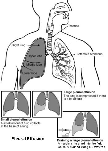
ومع ذلك، في العديد من الحالات، ما لم يتم علاج السبب الأساسي، فمن المحتمل أن يعود الانصباب خلال بضعة أسابيع. تصريف السائل بشكل متكرر عندما تصبح الأعراض مزعجة هو أحد الخيارات.
اعتمادًا على السبب الأساسي، تشمل خيارات العلاج الأخرى التي يتم النظر فيها أحيانًا:
Pleurodesis.
في هذا الإجراء، يتم حقن مادة كيميائية خاصة (مصلب) في الفراغ الجنبي. يتسبب ذلك في التهاب الأغشية الجنبية ويساعدها على 'الالتصاق' ببعضها البعض. يساعد ذلك في منع تراكم السوائل مرة أخرى في الانصباب. غالبًا ما يُستخدم التصاق الجنبة في علاج الانصبابات المتكررة الناتجة عن السرطان.
غالبًا ما يتم استخدام التصاق الجنبة بالتلك. إذا أعيد نفخ الرئة بعد تصريف السائل، يمكن استخدام مسحوق التلك المعقم للمساعدة في لصق الجنبة معًا. يضع الطبيب التلك عبر الأنبوب المتصل بالتصريف ثم يترك التصريف مغلقًا لمدة ساعة تقريبًا. هذا يتيح الوقت لبطانتي الرئة للالتصاق معًا. قد يقوم الطبيب بتوصيل التصريف بجهاز شفط لتطبيق كمية صغيرة من الضغط. يمكن أن يساعد هذا في إحكام إغلاق الجنبة معًا. بعد التصاق الجنبة، عادة ما يبقى التصريف في مكانه لمدة 24 ساعة أخرى.
مصارف دائمة
إذا كان الانصباب متكررًا (ربما لأن السبب الأساسي غير قابل للعلاج)، فمن الممكن وضع مصارف صدرية متكررة. قد يكون هذا غير مريح ويعني قضاء الكثير من الوقت في الدخول والخروج من المستشفى. في هذه الحالة، يمكن استخدام قسطرة خاصة يسمى قسطرة جنبية داخلية نفقية (TIPC)، يمكن أن تكون وضع. هذا يسمح بتصريف الانصباب الجنبي بسهولة أثناء وجودك في المنزل. عندما تشعر بتراكم السوائل على الرئة، يسمح القسطرة بتصريف السوائل في زجاجة. يمكن القيام بذلك من قبلك أو من قبل أحد أفراد العائلة أو ممرضة. يتم إدخال القسطرة في المستشفى في جناح نهاري. طالما لا توجد مضاعفات، يمكنك عادة العودة إلى المنزل في نفس اليوم.
إدخال تحويلة
عملية لإدخال تحويلة (مثل مصرف داخلي) للسماح بتصريف السوائل من الصدر إلى تجويف البطن. يُطلق على هذا اسم "تحويلة جنبية بطنية". نادرًا ما يتم استخدامها.
جراحة الصدر بمساعدة الفيديو
قد يكون من الممكن تصريف الانصباب الجنبي وإجراء التصاق الجنبة باستخدام إجراء يسمى تنظير الصدر. هذا نوع من الجراحة بالمنظار حيث يضع الطبيب أنبوبًا صغيرًا مرنًا (منظار الصدر) في الصدر. يحتوي الأنبوب على ضوء وكاميرا في نهايته، حتى يتمكن الأطباء من رؤية داخل صدرك. ولهذا السبب يُطلق عليه "مدعوم بالفيديو". يمكن استخدامه لأخذ خزعة، أو لتصريف الانصباب أو التصاق الجنبة. في هذا الإجراء، تستلقي على جانبك ويتم إعطاؤك حقنة من مهدئ لتشعر بالنعاس. يتم استخدام مخدر موضعي لتخدير المنطقة ويقوم الطبيب بعمل قطع صغير لوضع منظار الصدر. يستغرق الإجراء حوالي 40 إلى 60 دقيقة وبعد ذلك سيتم ترك أنبوب تصريف في الصدر لتصريف أي سوائل متبقية من تجويف الصدر.
تشمل الجراحة أيضًا عملية لإزالة الغشاء البلوري تُسمى استئصال الغشاء البلوري. تُستخدم أحيانًا في الأشخاص الذين يعانون من انصبابات بسبب السرطان عندما تفشل الخيارات العلاجية الأخرى.
اختيارات المرضى لـ حالات الرئة

الصدر والرئتين
التليف الرئوي
التليف الرئوي هو مرض رئوي خطير حيث تتضرر الأكياس الهوائية الصغيرة في الرئتين (الحويصلات الهوائية) والأنسجة الرئوية المجاورة للحويصلات وتصاب بالتندب، مما يؤدي إلى تليف الرئة. العرض الرئيسي هو ضيق التنفس الذي يزداد سوءًا تدريجيًا. غالبًا ما يكون السبب غير معروف، ولكن يمكن أن يحدث التليف الرئوي بسبب مجموعة متنوعة من الحالات الأساسية. تشمل العلاجات معالجة أي سبب أساسي بالإضافة إلى الستيرويدات والأدوية الأخرى. قد يُنظر بشكل متزايد في زراعة الرئة.
بقلم الدكتورة توني هازيل، FRCGP
الصدر والرئتين
تسمم بأول أكسيد الكربون
أول أكسيد الكربون هو غاز سام عديم الرائحة. حتى الكميات الصغيرة يمكن أن تحرم الجسم من الأكسجين، ويمكن أن تؤدي إلى تلف الدماغ في الحالات الشديدة. تصف هذه النشرة أعراض أول أكسيد الكربون وتقدم نصائح حول كيفية حماية نفسك من التسمم بأول أكسيد الكربون.
بقلم الدكتورة فيليبا فينسنت، MRCGP
الأسئلة الشائعة
كم من الوقت يمكن أن يستمر الانصباب الجنبي إذا تُرك دون علاج؟
تختلف مدة الانصباب الجنبي بشكل كبير اعتمادًا على السبب الأساسي له. قد تُترك الانصبابات الصغيرة التي لا تسبب أعراضًا وتُراقب، وقد تُشفى من تلقاء نفسها. ومع ذلك، إذا لم يتم علاج الحالة الأساسية، فمن المحتمل أن تعود الانصبابات الأكبر أو تستمر.
ما الفرق بين شفط السوائل الجنبيّة والتصاق الجنبة؟
شفط السوائل الجنبيّة (أو بزل الجنبة) هو إجراء لتصريف السوائل من الصدر باستخدام إبرة أو أنبوب، مما يوفر راحة فورية من الأعراض مثل ضيق التنفس. من ناحية أخرى، فإن التصاق الجنبة هو إجراء يتم فيه حقن مادة كيميائية خاصة في الفراغ الجنبي لجعل طبقتي الجنبة تلتصقان ببعضهما البعض، مما يمنع تراكم السوائل مرة أخرى. يركز الشفط على إزالة السوائل الموجودة، بينما يهدف التصاق الجنبة إلى منع تكرارها.
ما هو القسطار الجنبي الدائم (TIPC) وكيف يساعد؟
القسطرة الجنابية الدائمة (TIPC) هي أنبوب تصريف خاص يمكن إدخاله لإدارة الانصباب الجنبي المتكرر. يسمح بتصريف السوائل بسهولة في المنزل بواسطة المريض أو أحد أفراد العائلة أو ممرضة، دون الحاجة إلى زيارات متكررة للمستشفى للتصريف. يساعد ذلك في إدارة الأعراض عندما لا يمكن علاج السبب الأساسي للانصباب بشكل كامل.
متى يمكن اعتبار الجراحة لعلاج الانصباب الجنبي بخلاف التصريف؟
بجانب تصريف السوائل، قد يُنظر في الجراحة للحالات المتكررة أو لأسباب محددة. تشمل الخيارات الجراحة الصدرية بمساعدة الفيديو (VATS) لتصريف السوائل، إجراء التصاق الجنبة، أو أخذ خزعة. في حالات الانصباب المتكرر، يمكن إدخال تحويلة جنبية بطنية لتصريف السوائل إلى تجويف البطن. نادرًا، قد تُستخدم عملية لإزالة الجنبة، تُسمى استئصال الجنبة، للانصبابات المرتبطة بالسرطان عندما تفشل العلاجات الأخرى.
هل يمكن علاج الانصباب الجنبي بشكل كامل؟
يعتمد ما إذا كان يمكن علاج الانصباب الجنبي بشكل كامل على ما إذا كان يمكن علاج السبب الأساسي بنجاح. إذا تم معالجة السبب الجذري، مثل عدوى الرئة، بشكل فعال، فهناك فرصة جيدة أن يزول الانصباب الجنبي بشكل دائم. ومع ذلك، إذا لم يكن من الممكن علاج الحالة الأساسية، أو تم علاجها جزئيًا فقط، فقد يتكرر الانصباب حتى بعد التصريف.
قراءة إضافية ومراجع
- إرشادات الجمعية البريطانية لأمراض الصدر لأمراض الجنبة; الجمعية البريطانية لأمراض الصدر - BMJ (2023).
- كريشنا ر، رودرابا م; الانصباب الجنبي. ستات بيرلز 2020.
- Skok K, Hladnik G, Grm A, et al; الانصباب الجنبي الخبيث وإدارته الحالية: مراجعة. ميديسينا (كاوناس). 15 أغسطس 2019;55(8). معرف الوثيقة: medicina55080490. doi: 10.3390/medicina55080490.
- Bibby AC, Dorn P, Psallidas I, et al; بيان ERS/EACTS حول إدارة الانصباب الجنبي الخبيث. Eur Respir J. 27 يوليو 2018;52(1). pii: 13993003.00349-2018. doi: 10.1183/13993003.00349-2018. طباعة يوليو 2018.
- Kulandaisamy PC, Kulandaisamy S, Kramer D, et al; الانصباب الجنبي الخبيث - مراجعة للمبادئ التوجيهية والممارسات الحالية. مجلة الطب السريري. 26 نوفمبر 2021;10(23). معرف الوثيقة: jcm10235535. doi: 10.3390/jcm10235535.
عن المؤلفعرض السيرة الذاتية الكاملة

الدكتورة هايلي ويلاسي، زميلة الكلية الملكية للأطباء العامين
طبيب عام، مؤلف طبي
MBChB (1992), DRCOG, DFFP, MRCOG (Part 1) MRCGP (2007), DFSRH (2013), MSc - medical education (2020)
كانت الدكتورة هايلي ويلاسي طبيبة عامة في هيئة الخدمات الصحية الوطنية تعمل في شمال غرب إنجلترا، وتقاعدت من الممارسة السريرية في عام 2022 بعد 30 عامًا.
حول المراجععرض السيرة الذاتية الكاملة

الدكتور دوغ مكيتشني، MRCGP
كاتب طبي
MA, MBBS, MSc, DRCOG, MRCP(UK), MRCGP(2021), FHEA
الدكتور دوغ مكيتشني هو طبيب عام في هيئة الخدمات الصحية الوطنية يعمل في لندن. يعمل بدوام كامل في المجال السريري وهو أيضًا نائب رئيس وحدة الممارسة السريرية والمهنية في كلية الطب بجامعة كوليدج لندن.
تاريخ المقال
تمت كتابة المعلومات على هذه الصفحة ومراجعتها من قبل أطباء مؤهلين.
المقال متاح أيضًا باللغة الإنجليزية, الألمانية, إسبانية, الفرنسية, إيطالي, البرتغالية, الهندية, العبرية, العربية ,، و السويدية.
المراجعة التالية مستحقة: 19 ديسمبر 2028
21 ديسمبر 2023 | أحدث إصدار
آخر تحديث بواسطة
الدكتورة هايلي ويلاسي، زميلة الكلية الملكية للأطباء العامينمراجعة من قبل
الدكتور دوغ مكيتشني، MRCGP

اسأل، شارك، تواصل.
تصفح المناقشات، اطرح الأسئلة، وشارك التجارب عبر مئات المواضيع الصحية.

هل تشعر بتوعك؟
قم بتقييم أعراضك عبر الإنترنت مجانًا
اشترك في النشرة الإخبارية للمرضى
جرعتك الأسبوعية من النصائح الصحية الواضحة والموثوقة - مكتوبة لمساعدتك على الشعور بالاطلاع والثقة والتحكم.
من خلال الاشتراك، فإنك تقبل سياسة الخصوصية. يمكنك إلغاء الاشتراك في أي وقت. نحن لا نبيع بياناتك أبدًا.
المزيد في الصدر والرئتين
- التفاقمات الحادة لمرض الانسداد الرئوي المزمن
- الذبحة الصدرية
- الالتهاب الرئوي الشفطي
- التهاب القصيبات
- تسمم بأول أكسيد الكربون
- التهاب الغضروف الضلعي
- كوفيد-19
- التهاب لسان المزمار
- سرطان الرئة
- ورم الظهارة المتوسطة
- الذبحة الصدرية الدقيقة
- موسعات الشعب الهوائية الفموية
- التهاب الجنبة
- الالتهاب الرئوي
- استرواح الصدر
- التليف الرئوي
- الوذمة الرئوية
- الساركويد
- فيروسات سارس وميرس
- السعال الديكي