Mitral stenosis
Peer reviewed by Dr Adrian Bonsall, MBBSLast updated by Dr Colin Tidy, MRCGPLast updated 1 Aug 2017
Meets Patient’s editorial guidelines
- DownloadDownload
- Share
- Language
- Discussion
- Audio Version
- Add to preferred sources on Google
In this series:Heart valve diseaseMitral regurgitationAortic stenosisAortic regurgitationInfective endocarditis
Content notice
This page hasn’t been reviewed recently, so some information or external links may have changed.
Mitral stenosis means that when the mitral valve opens, it does not open fully. The opening is therefore narrower than normal (stenosed).
At a glance
Mitral stenosis is a condition where the heart's mitral valve does not open fully.
This restricts blood flow from the left atrium to the left ventricle.
Symptoms can include shortness of breath, tiredness, swollen ankles, and chest pain.
Mild cases may not need treatment, but medication can help ease symptoms.
More severe cases may require surgery to widen or replace the valve.
Editor’s note
Dr Sarah Jarvis, 27th November 2021
NICE guidance on heart valve disease
This leaflet has been archived and has not been reviewed since 2017. It does not include the latest guidance on investigations and treatments for mitral stenosis, which were produced by the National Institute for Health and Care Excellence (NICE) in November 2021.
You can find out more about NICE's recommendations from the further reading section at the end of this leaflet.
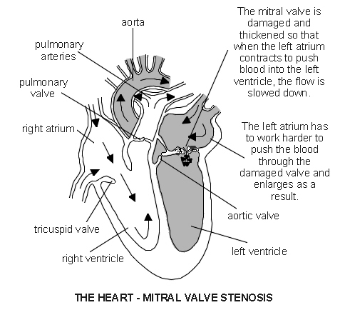
The mitral valve is a heart valve that lies between the left atrium and left ventricle. The valve has two flaps (cusps). The valve allows blood to flow into the left ventricle when the left atrium squeezes (contracts). When the left ventricle contracts, the valve closes and the blood flows out through the aortic valve into the aorta. (The aorta is the main artery which takes blood to the body.)
The cusps are stopped from turning inside out by thin strands of tissue called chordae. The chordae anchor the cusps to the inside wall of the ventricle. The valve or chordae may get damaged or scarred which can prevent the valve from working properly. This can lead to disorders called mitral stenosis, mitral regurgitation, or both.
Mitral stenosis means that when the mitral valve opens, it does not open fully. The opening is therefore narrower than normal (stenosed). So, there is some restriction of blood flow from the left atrium to the left ventricle. This in turn means there is a reduced amount of blood that is pumped out into the body from the left ventricle. In general, the more narrowed the valve, the less blood can get through and the more severe the problem is likely to be.
Symptoms
If the valve is only mildly narrowed (stenosed) you may have no symptoms or problems. If the stenosis is more severe, the symptoms may include:
Shortness of breath, especially with activity or when you lie down
Swollen ankles (oedema)
The heart - mitral valve stenosis
Treatment
Medication
Mild cases may not require any regular medication. Although medicines cannot correct a narrowed (stenosed) mitral valve, some medicines may be prescribed to help ease symptoms, or to help prevent complications - for example, angiotensin-converting enzyme (ACE) inhibitors, 'water tablets' (diuretics) and anticoagulation medication. If you develop atrial fibrillation, several medicines can be used to slow the heart rate down.
Surgical treatment
Surgical treatment is needed in more severe cases. There are various options, depending of the exact site and severity of the stenosis.
Stretching the stenosed valve
This is a procedure that does not involve open heart surgery. It is called percutaneous balloon commissurotomy or balloon valvuloplasty. (It is called a commissurotomy, as the area where the valve flaps (cusps) come into contact with each other are known as the commissures.)
It is done by inserting a thin tube called a catheter through the skin (percutaneous) into the main blood vessel in the top of the leg. The catheter is passed up to the heart. The tip of the catheter is placed in the mitral valve opening. A balloon at the tip of the catheter is then inflated to stretch the narrowed valve. This is often successful in widening the narrowed valve.
Valve repair is possible in some cases
This is called mitral commissurotomy or mitral valvotomy. This is usually done by open heart surgery. Basically, the edges (commissures) of valve cusps that have become scarred and fused are shaved back to widen the narrowed valve opening.
Valve replacement is needed in some cases
This may be with a mechanical or a tissue valve.
Mechanical valves are made of materials which are not likely to react with your body (for example, those made from titanium), although they can produce a noise which can be heard outside the body.
Tissue valves are made from treated animal tissue (for example, valves from a pig).
If you need surgery, a surgeon will advise on which is the best option for your situation.
What is the outlook for people with mitral stenosis?
In some cases, the disorder is mild and causes no symptoms. If you develop symptoms they tend to become gradually worse over the years. However, the speed of decline can vary. It often takes years for symptoms to become serious. Medication can ease symptoms but cannot reverse a narrowed (stenosed) valve.
Surgical treatments have greatly improved the outlook (prognosis) for most people with more severe stenosis. Surgery has a very good success rate. However, as with all surgical procedures and operations, there is some risk involved when you have surgery. Complications due to surgery occur in a small number of cases.
Patient picks for Heart disease
Heart health and blood vessels
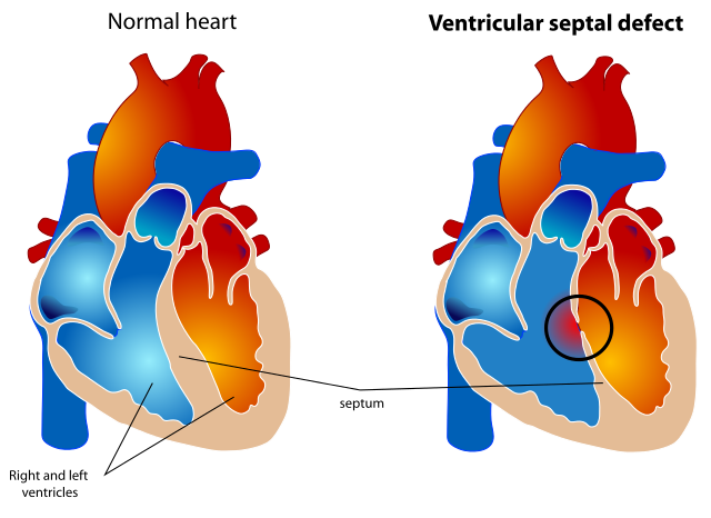
Ventricular septal defect
Ventricular septal defect (VSD) is a gap or defect in the septum between the heart's two lower chambers (ventricles). The septum is a wall that separates the heart's right and left side of the heart. Septal defects are sometimes called a 'hole' in the heart. It is the most common heart problem that babies are born with. Many defects in the ventricular septum close themselves and cause no problems. Otherwise, medicines or surgery can help. Most babies born with a defect in the septum have normal survival.
by Dr Hayley Willacy, FRCGP
Heart health and blood vessels
Microvascular angina
There is no agreed definition for CSX. The underlying cause may be sudden narrowing (spasm) of normal coronary arteries without any evidence of atheromatous fatty plaques.
by Dr Hayley Willacy, FRCGP
Frequently asked questions
What is the mitral valve's normal function in the heart?
The mitral valve is located between the left atrium and the left ventricle. Its purpose is to allow blood to flow from the left atrium into the left ventricle when the atrium contracts. When the left ventricle then contracts, the valve closes, directing blood out through the aortic valve to the rest of the body.
How do the chordae help the mitral valve work correctly?
The chordae are thin strands of tissue that are attached to the mitral valve's flaps (cusps) and anchor them to the inner wall of the left ventricle. Their role is crucial in preventing the valve cusps from turning inside out when the heart contracts, ensuring proper blood flow control.
What are the differences between mechanical and tissue valves used for replacement?
Mechanical valves are made from durable materials like titanium, which are designed not to react with the body, although they can sometimes produce an audible sound. Tissue valves, on the other hand, are made from treated animal tissue, such as from pigs.
What happens if the mitral valve or chordae get damaged?
Damage or scarring to the mitral valve or its chordae can prevent the valve from functioning correctly. This can lead to conditions such as mitral stenosis, where the valve doesn't open fully, or mitral regurgitation, or both, disrupting normal blood flow through the heart.
Further reading and references
- Prophylaxis against infective endocarditis: Antimicrobial prophylaxis against infective endocarditis in adults and children undergoing interventional procedures; NICE Clinical Guideline (March 2008 - last updated July 2016)
- 2023 ESC Guidelines for the management of infective endocarditis; European Society of Cardiology (Aug 2023)
- Nishimura RA, Otto CM, Bonow RO, et al; 2017 AHA/ACC Focused Update of the 2014 AHA/ACC Guideline for the Management of Patients With Valvular Heart Disease. Circulation. 2017; CIR.0000000000000503. Originally published March 15, 2017.
- Vahanian A et al; Guidelines on the management of valvular heart disease: The Task Force on the Management of Valvular Heart Disease of the European Society of Cardiology, 2017
- Ozkan M; What is new in ACC/AHA 2017 focused update of valvular heart disease guidelines. Anatol J Cardiol. 2017 Jun;17(6):421-422. doi: 10.14744/AnatolJCardiol.2017.7925.
- Heart valve disease presenting in adults: investigation and management; NICE guideline (November 2021)
About the authorView full bio
Updated by

Dr Colin Tidy, MRCGP
General Practitioner, Medical Author
MBBS, MRCGP, MRCP (Paediatrics), DCH
Dr Colin Tidy is an NHS Doctor, based in Oxfordshire.
About the reviewerView full bio

Dr Adrian Bonsall, MBBS
Medical Author
MA (Chemistry), MBBS (Hons), DCH
Since 2000 Adrian has been employed in emergency and critical care paediatrics based in Sydney, with particular interests in toxicology, trauma and resuscitation.
Join the conversation
Share your thoughts or experiences with others in our patient communities.

Ask, share, connect.
Browse discussions, ask questions, and share experiences across hundreds of health topics.

Feeling unwell?
Assess your symptoms online for free
Sign up to the Patient newsletter
Your weekly dose of clear, trustworthy health advice - written to help you feel informed, confident and in control.
By subscribing you accept our Privacy Policy. You can unsubscribe at any time. We never sell your data.