Cystoscopy
مراجعة من قبل الدكتور كولين تايدي، MRCGPآخر تحديث بواسطة الدكتورة فيليبا فينسنت، MRCGPLast updated 10 Aug 2023
يتوافق مع الإرشادات التحريرية
- تنزيلتنزيل
- مشاركة
- Language
- نقاش
- نسخة صوتية
- Add to preferred sources on Google
A cystoscopy is a procedure which looks into the bladder with a special telescope called a cystoscope.
ملاحظة: المعلومات أدناه هي دليل عام فقط. قد تختلف الترتيبات وطريقة إجراء الاختبارات بين المستشفيات المختلفة. دائماً اتبع التعليمات المقدمة من طبيبك أو المستشفى المحلي.
At a glance
A cystoscopy uses a thin telescope to look inside the bladder via the urethra.
It can diagnose frequent urine infections, blood in urine, or persistent pain when urinating.
A cystoscopy can also treat conditions like bladder stones or small bladder tumours.
The procedure usually takes 5-10 minutes and is often done while you are awake.
You might feel a mild burning or frequent need to urinate for a day after.
Seek medical help if pain or bleeding is severe or lasts over two days, or if you get a fever.
في هذه المقالة:
Video picks for اختبارات البول والمثانة
تابع القراءة أدناه
Why is a cystoscopy done?
To help with diagnosis
A cystoscopy may be done to help to find the cause of symptoms such as:
Unusual cells found in a urine sample.
Persistent pain when passing urine.
Difficulty in passing urine - which may be due to prostate enlargement or a narrowing (stricture) of the urethra.
Cystoscopy may also be done to monitor progress of conditions. For example, some people have a routine cystoscopy every now and then after treatment for a bladder tumour. This helps to detect any early recurrence which can be treated before it spreads further.
To treat certain conditions, or to do certain procedures
By using various instruments which are passed down the side channels a doctor can:
Remove bladder stones. If a stone is lodged higher - in a ureter - the doctor may extend the cystoscope up into a ureter. The ureters are the tubes that drain urine from the kidneys to the bladder.
Remove small lumps (polyps) or الأورام from the lining of the bladder.
Insert a small tube (stent) into a narrowed ureter. This helps the flow of urine if there is a narrowing.
Perform a special X-ray of the ureters and kidneys. A doctor can inject a dye into the ureters up towards the kidney. This shows up on X-ray pictures and helps to show problems of the kidney or ureter.
Remove the prostate gland (by using a special type of cystoscope which 'chips' away at the prostate gland bit by bit).
What is a cystoscope?
العودة إلى المحتوياتA cystoscope is a thin telescope which is passed into the bladder via the urethra, the tube that takes urine from the bladder to the outside of the body. There are two types. The cystoscope can be looked down but it also transmits pictures to a computer screen so they can be seen by other members of the team. Both types of cystoscope have side channels where various thin instruments can be passed down. For example a small sample (خزعة) may be taken from the bladder wall using an instrument which can pinch tissue and carry it back down the side channel.
تابع القراءة أدناه
Types of cystoscopy
العودة إلى المحتوياتA flexible cystoscope is the most commonly used. It is a thin, flexible, fibre-optic telescope which is about as thick as a pencil. As it is flexible, it passes easily along the curves of the urethra. The flexible tip can also be moved easily so that the whole of the bladder lining can be seen.
Flexible cystoscope
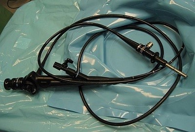
© By Michael Reeve, via Wikimedia Commons
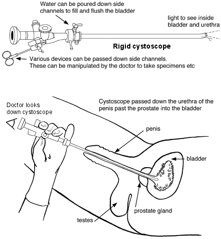
A rigid cystoscope is a thin, solid, straight telescope. It allows a greater variety of devices to pass down the side channels so can be used for a wider variety of procedures.
Diagram of a rigid cystoscope and how a cystoscopy is performed
Cystoscopy procedure
العودة إلى المحتوياتCystoscopy is usually done as an outpatient procedure. This means people go home the same day and aren't required to stay overnight. It is usually done whilst awake. Some people are given a sedative to help them to relax.
The opening to the urethra (at the end of the penis, or the outside of the vagina) and the nearby skin will be cleaned. Some 'jelly' is then squirted into the opening of the urethra. The jelly usually contains a local anaesthetic to numb the lining of the urethra. This helps the cystoscope to pass into the urethra with as little discomfort as possible.
Male and female cystoscopy
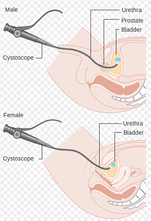
The cystoscope is then gently pushed up into the bladder and the lining of the urethra and bladder is carefully examined. Sterile water is passed down a side channel in the cystoscope to fill the bladder slowly. This makes it easier to see the lining of the bladder and the bladder wall. As the bladder fills it causes the feeling of a need to pass urine which may be uncomfortable.
The cystoscope is then gently pulled out. If a biopsy was taken, the sample would be sent away to be tested and looked at under a microscope. It can take several weeks for the report of the biopsy to come back to the doctor.
In some cases a general anaesthetic is given when a cystoscopy is done, particularly if a rigid cystoscope is used. In some cases a spinal anaesthetic is given which numbs all the lower half of the body.
تابع القراءة أدناه
How long does a cystoscopy take?
العودة إلى المحتوياتA cystoscopy takes about 5-10 minutes if it is just to look inside the bladder. It may last longer if a procedure is being performed - for example, taking a sample (خزعة) from the lining of the bladder.
Is a cystoscopy painful?
العودة إلى المحتوياتCystoscopies are not usually painful, particularly as local anaesthetic is used to insert the cystoscope. They can be uncomfortable, especially when the bladder has been filled with water.
Complications of a cystoscopy
العودة إلى المحتوياتMost cystoscopies are done without any problem.
For the following 24 hours there may be a mild burning sensation when passing urine and a feeling of a need to pass urine more often than usual. The urine may also look pink due to mild bleeding, particularly if a biopsy was taken.
Occasionally, a عدوى بولية develops shortly after a cystoscopy. This can cause a ارتفاع درجة الحرارة (الحمى) and pain on passing urine.
Rarely, the cystoscope may damage or perforate the bladder. After having a cystoscopy, medical advice should be sought if:
Pain or bleeding is severe.
Pain or bleeding lasts longer than two days.
Symptoms of infection develop, such as a fever.
Preparing for a cystoscopy
العودة إلى المحتوياتThere are not usually any particular preparations needed before a flexible cystoscopy. Usually it is possible to eat and drink as normal beforehand. There is no need to empty the bladder. A rigid cystoscopy normally requires a general anaesthetic so guidance will be given about when to stop eating and drinking and what to do.
After a cystoscopy
العودة إلى المحتوياتAfter a flexible cystoscopy, it is usual to need to empty the bladder urgently and then to go home without any need for additional support. After a rigid cystoscopy, a catheter may be used to empty the bladder and the normal advice would be given about having a general anaesthetic (to wait in the hospital for a few hours to recover, to be unable to drive home, to need support at home for 24 hours).
Patient picks for اختبارات البول والمثانة
الفحوصات والتحقيقات
Ketones in urine
Ketones are produced when the body burns fat for fuel. Normally these ketones will be completely broken down (metabolised) so that there are very few ketones in the urine. If for any reason the body cannot get enough glucose for energy it will switch to using body fats, causing an increase in ketones in the body. This results in more ketones in urine.
بواسطة الدكتورة سورانجي مينديس، MRCGP

الفحوصات والتحقيقات
Urodynamic testing
Urodynamic tests check the filling and emptying of your bladder and help to investigate the cause of any urinary incontinence you may have. Note: the information below is a general guide only. The arrangements, and the way tests are performed, may vary between different hospitals. Always follow the instructions given by your doctor or local hospital.
by Dr Hayley Willacy, FRCGP
الأسئلة الشائعة
What is the urethra?
The urethra is the tube that carries urine from the bladder to the outside of the body. A cystoscope is passed into the bladder through this tube during the procedure.
How is a biopsy taken during a cystoscopy, and how long until I get the results?
During a cystoscopy, a small sample (biopsy) can be taken from the bladder wall using an instrument passed down a side channel of the cystoscope. This instrument can pinch tissue and carry it back. After the sample is taken, it's sent for testing and examined under a microscope. It can take several weeks for the report of the biopsy to be sent back to your doctor.
Will I be given anything to help with pain during the cystoscopy?
Yes, a local anaesthetic jelly is usually squirted into the opening of the urethra before the cystoscope is inserted. This numbs the lining of the urethra to help the cystoscope pass with as little discomfort as possible. Some people may also be given a sedative to help them relax during the procedure.
What should I do if my symptoms like burning or bleeding continue for more than two days after the procedure?
You should seek medical advice if pain or bleeding is severe or lasts longer than two days after a cystoscopy. It's also important to contact a doctor if you develop symptoms of an infection, such as a fever.
How do I prepare for a flexible cystoscopy compared to a rigid one?
For a flexible cystoscopy, you typically don't need any special preparations and can eat and drink as normal beforehand, with no need to empty your bladder. However, a rigid cystoscopy usually requires a general anaesthetic, so you would receive specific instructions on when to stop eating and drinking and what else you need to do to prepare.
What happens immediately after a flexible cystoscopy?
After a flexible cystoscopy, it's common to feel an urgent need to empty your bladder. You can usually go home without needing additional support. For rigid cystoscopies, different advice applies due to the general anaesthetic.
قراءة إضافية ومراجع
- Zhang ZS, Wang XL, Xu CL, et al; Music reduces panic: an initial study of listening to preferred music improves male patient discomfort and anxiety during flexible cystoscopy. J Endourol. 2014 Jun;28(6):739-44. doi: 10.1089/end.2013.0705. Epub 2014 Mar 31.
- Gee JR, Waterman BJ, Jarrard DF, et al; Flexible and rigid cystoscopy in women. JSLS. 2009 Apr-Jun;13(2):135-8.
- Engelsgjerd JS, Deibert CM; Cystoscopy
- DeGeorge KC, Holt HR, Hodges SC; Bladder Cancer: Diagnosis and Treatment. Am Fam Physician. 2017 Oct 15;96(8):507-514.
تابع القراءة أدناه
About the authorView full bio

الدكتورة فيليبا فينسنت، MRCGP
General Practitioner, Medical Author
MB BS, Bsc, MRCGP (2000), DCH, DFSRH, DRCOG
الدكتورة Philippa Vincent is an NHS GP working in North London.
About the reviewerView full bio

الدكتور كولين تايدي، MRCGP
General Practitioner, Medical Author
MBBS, MRCGP, MRCP (Paediatrics), DCH
Dr Colin Tidy is an NHS Doctor, based in Oxfordshire.
تاريخ المقال
تمت كتابة المعلومات على هذه الصفحة ومراجعتها من قبل أطباء مؤهلين.
Next review due: 8 Aug 2028
10 Aug 2023 | أحدث إصدار

اسأل، شارك، تواصل.
تصفح المناقشات، اطرح الأسئلة، وشارك التجارب عبر مئات المواضيع الصحية.

هل تشعر بتوعك؟
قم بتقييم أعراضك عبر الإنترنت مجانًا
اشترك في النشرة الإخبارية للمرضى
جرعتك الأسبوعية من النصائح الصحية الواضحة والموثوقة - مكتوبة لمساعدتك على الشعور بالاطلاع والثقة والتحكم.
By subscribing you accept our سياسة الخصوصية. يمكنك إلغاء الاشتراك في أي وقت. نحن لا نبيع بياناتك أبدًا.